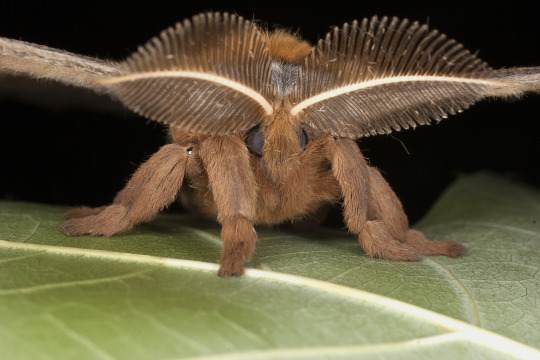
“A picture tells 1,000 words.”
Specimen imaging is a method of documenting specimens in the Invertebrate Zoology collection for research as well as collection maintenance. Digitizing the collection allows it to be more accessible to the scientific community. Specimen-based documentation includes capturing the data from the specimen as well as images of the specimen from different angles. Images of labels serve as a primary data capture that may then be used to populate a database of specimen records.
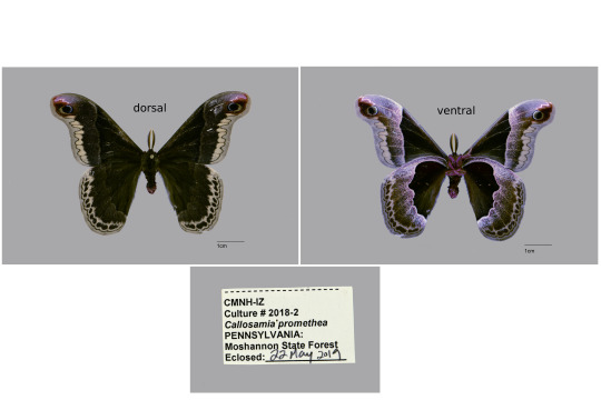
Most of the imaging currently being done is based on requests from the entomology community needing images of specimens known to be deposited at the Carnegie. There are many historical specimens that are not otherwise imaged but are referenced in older publications. These specimens include cataloged species vouchers referred to as types, or specimens referenced in publications that are of interest to researchers studying those species.
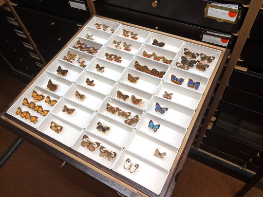
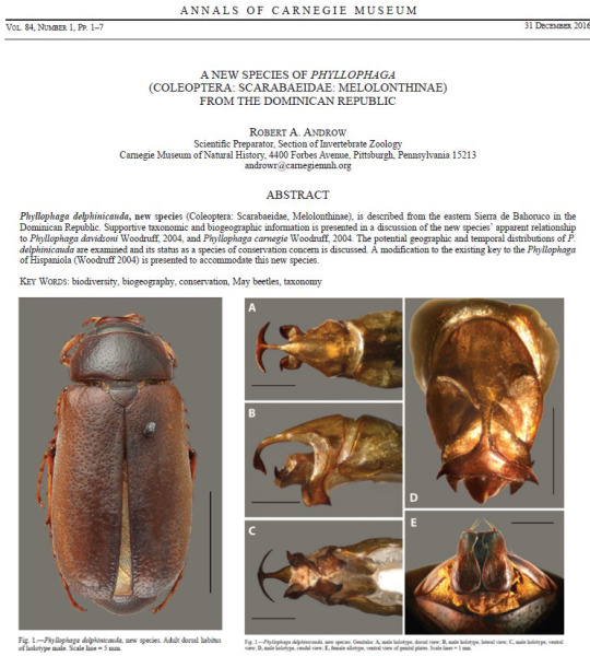
Specimen photography is also essential when discovering new species. When a paper is published describing a new species, images of the designated types are included. The type series includes the series of specimens that were examined and used to describe the new species in detail. These images should be taken with a scale line to show the size of the specimen. Images of prepared dissections are also included.
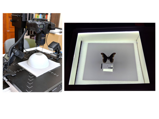
Imaging techniques include using a copy stand, flash lighting, and focus-stacking through software that produces a final high-quality image with all parts of the specimen in focus. A light box may also be used, as an alternative to flash lighting, to provide even lighting and sharp images.
A macro ring flash helps with producing even lighting when imaging live insects that are moving or are very small and need to be really close to the lens.
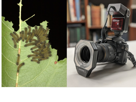
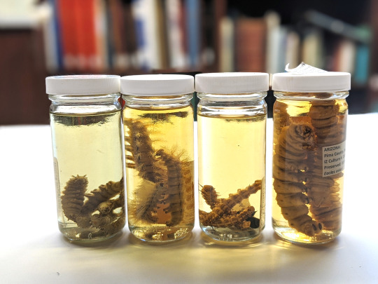
Photographing live specimens that will lose their color when preserved in alcohol, such as caterpillars, is crucial. Larval images are a major component of the caterpillar collection and are incredibly valuable documentation of larval growth. Raising caterpillars is a way to document the life history of different species of moths and butterflies, and their associated caterpillars. Since the caterpillars will be stored in alcohol, the color will be lost in the preserved specimen, but these characteristics will be recorded through high-quality images.
Several other types of equipment are used to capture images at higher magnification so that characters may be seen in greater detail. Taking an image through a compound microscope allows one to capture an image that may be used to draw a detailed illustration. An image taken through a Scanning Electron Microscope offers even greater detail. All-together, the image collection is a major component of the archived data that contributes to the understanding of the specimens in the collection. This includes digital files and images on older slide film that still need to be scanned. The digital image collection continues to grow daily, and serves the broader entomological community that needs access to the reference specimens stored in the Invertebrate Zoology collection at the Carnegie.
Vanessa Verdecia is a collection assistant in the museum’s Invertebrate Zoology Section. Museum employees are encouraged to blog about their unique experiences and knowledge gained from working at the museum.
