• Skip to primary navigation
  • Skip to main content
  • Skip to primary sidebar

Carnegie Museum of Natural History

One of the Four Carnegie Museums of Pittsburgh

  • Visit
    • Buy Tickets
    • Visitor Information
    • Exhibitions
    • Events
    • Dining at the Museum
    • Celebrate at the Museum
    • Powdermill Nature Reserve
    • Event Venue Rental
  • Learn
    • Field Trips
    • Educator Information
    • Programs at the Museum
    • Bring the Museum to You
    • Guided Programs FAQ
    • Programs Online
    • Climate and Rural Systems Partnership
  • Research
    • Scientific Sections
    • Science Stories
    • Science Videos
    • Senior Science & Research Staff
    • Museum Library
    • Science Seminars
    • Scientific Publications
    • Specimen and Artifact Identification
  • About
    • Mission & Commitments
    • Directors Team
    • Museum History
  • Tickets
  • Give
  • Shop

amphibians and reptiles

April 20, 2021 by wpengine

An Illuminating Tale of Tracking Turtles

by Amanda K. Martin

*All research was conducted under approved permits from by IACUC, ODNR, and Metroparks. Do not try this at home with local wildlife. Photos by A. Martin unless noted otherwise.

Where do eastern box turtles go? When I started my graduate schooling in Dr. Karen Root’s lab at Bowling Green State University in Ohio, I was quite intrigued by this question. To address it, I conducted a study of box turtle movements in the Oak Openings Region, the distinctive landscape of oak savannas, woodlands, and wet prairies that stretches across seven counties in Northwest Ohio and Southeast Michigan.

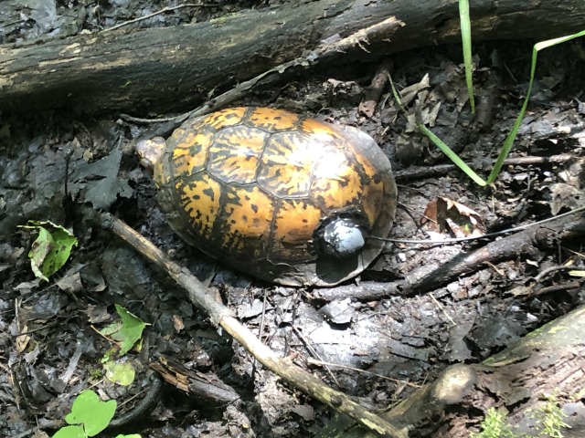
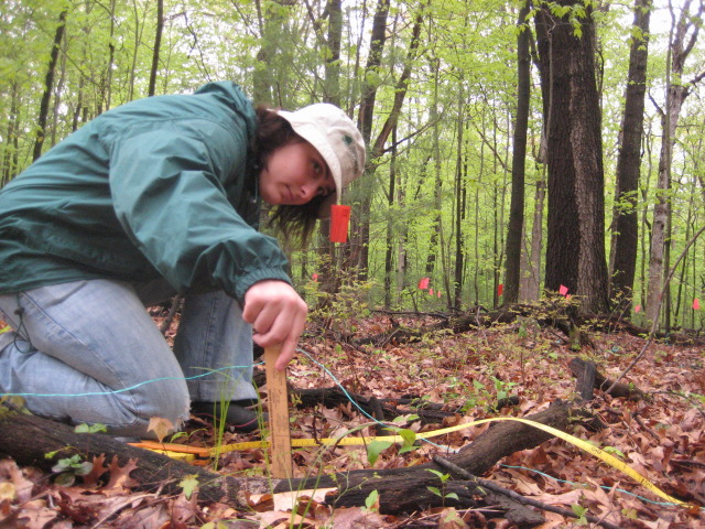
A method called radio telemetry was vital to my work. I walked around under the forest canopy searching for individuals (female or male) and whenever I found one, typically sitting still on the ground, I would pick it up while wearing gloves. In order to track its movements, I attached a radio transmitter onto the carapace (upper shell) using a special type of glue (Fig. 1A). After about a month of searching, I was able to track box turtles at two locations in the Toledo Metroparks system, six individuals in Oak Openings Preserve, and three individuals in Secor Metroparks.

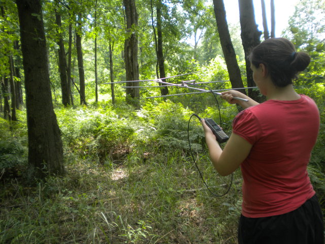
Two to three times a week, I would travel to these local parks and track each turtle using a silver three-pronged antenna and attached receiver. This portable combination detects the signal frequency produced by the transmitter on the tagged turtles, generating a “beeping” sound as it receives the electronic pulse. Guided by “beeps” I could re-find each turtle within an hour (Fig. 1B) depending on how dense the forest understory was. If I walked in the wrong direction, the noise would fade away and become quieter, but as I moved closer to the turtle’s location, the “beeping” sound would get louder and more frequent until I reached the turtle. Sometimes I would walk right past an individual sitting quietly in the leaf litter or under a log as their shell is often highly camouflaged to blend with the sunlit and shadowed patterns of a forest floor. One nice aspect of tracking box turtles with radio telemetry is that they do not run away very quickly, so they are easy to follow!

turtle on the ground among sticks and leaves
woman holding an antenna and receiver in the woods
Fig. 1A (top) and Fig. 1B (bottom): A box turtles with a transmitter (A) tracked by A. Martin using radio telemetry (antenna and receiver; B) in Oak Openings Region, Ohio, USA. Photo by S. Martin (B).

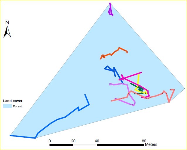
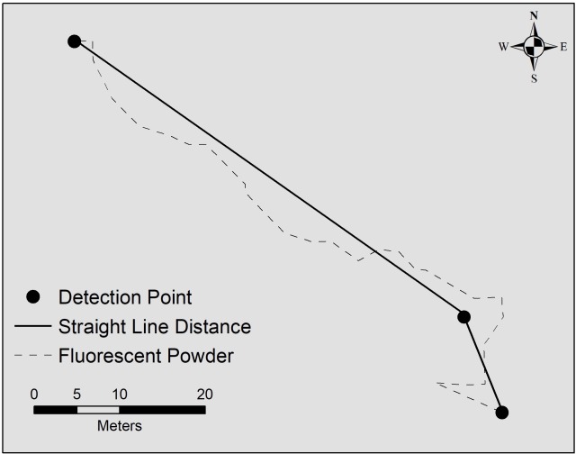
Radio telemetry is an excellent method for re-locating individuals, and provides a snapshot of where the individual is at a given time. With long-term tracking over the active season (mid-March to early November), researchers can better understand movements within a turtle’s home range, the area the animal regularly travels to meet its daily requirements, including food, shelter, and thermoregulation. Home ranges are estimated by drawing an outline around the outermost locations where a turtle was detected throughout the year, and assuming that the individual uses the area inside this boundary (Fig. 2A). Each time a turtle was found, I recorded the GPS coordinates of its location, and could then measure how far the turtle traveled by drawing a straight line between each location point. However, turtles may not always travel in a straight line, but rather follow an indirect route between detection points (Fig. 2B), so this method likely underestimates actual travel distance.

blue diagram showing box turtle home range
diagram showing box turtle distance traveled
Fig. 2A (top) and Fig. 2B (bottom): Box turtle home range (blue area) with daily movements (each color represents one day of travel) using fluorescent powder (A) and an example of an estimated distance traveled (solid black straight line) and actual distance traveled (dotted black curvier line) between location points (black circles; B).

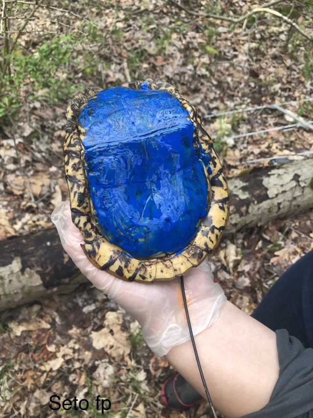
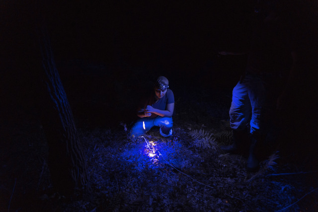
A research technique involving fluorescent powder can produce a far more accurate picture of daily box turtle movements. Non-toxic fluorescent powder is applied to the turtle’s plastron (underside; Fig. 3A) which then leaves a distinct trail as the turtle travels throughout its environment. At night, with the use of an ultraviolet light (Fig. 3B) these trails can then be illuminated, traced, and mapped. Since box turtles tend to travel near or over the same pathways, and because individual home ranges frequently overlap, multiple powder colors are required for some tracking studies.

I used multiple colors (red, blue, yellow, orange) for different days and individuals. The results of my tracking work using this technique demonstrated that box turtles traveled 32 meters per day, with females traveling slightly less than males, and that 95% of movements were less than 6 meters.

box turtle held in a person's hand
two people at night in the forest illuminated by blue light
woman with a ruler in the forest
Fig. 3A (top), Fig. 3B (middle), and Fig. 3C (bottom): A freshly painted plastron of a male box turtle (A), A. Martin with a field assistant illuminating the fluorescent powder trail with an ultraviolet light (B, photo by A. Kappler), and A. Martin measuring leaf litter along a box turtle’s pathway (C).

Tracking animals with fluorescent powder is more laborious than radio telemetry but demonstrates fine scale movement patterns not detected by radio telemetry. The frequent use of short movements, for example, is likely related to thermoregulation requirements (the need to move in and out of cool, shady patches), or encounters with multiple obstacles ranging from small to large logs, dense shrubs, and trees. Radio telemetry provides an estimation of home range size, while fluorescent powder tracking provides details on how that home range is utilized. In tandem, these research tools can provide important information on habitat use for local land managers, who can facilitate preservation of these reptiles.

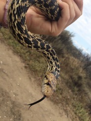
For more information on this project, including data on eastern garter snake movements, check out Chapter 4 of my dissertation.

Amanda K. Martin is a Post-doctoral Researcher in Section of Amphibians and Reptiles. Museum employees are encouraged to blog about their unique experiences and knowledge gained from working at the museum.

Related Content

Turtle Bottoms

A Wood Turtle at Powdermill

Ask a Scientist: How does climate change affect turtle behavior?

Carnegie Museum of Natural History Blog Citation Information

Blog author: Martin, Amanda K.
Publication date: April 20, 2021

Share this post!

  • Share on Twitter Share on Twitter
  • Share on Facebook Share on Facebook
  • Share on Pinterest Share on Pinterest
  • Share on LinkedIn Share on LinkedIn
  • Share on Reddit Share on Reddit
  • Share via Email Share via Email

Share this post!

  • Share on Twitter Share on Twitter
  • Share on Facebook Share on Facebook
  • Share on Pinterest Share on Pinterest
  • Share on LinkedIn Share on LinkedIn
  • Share on Reddit Share on Reddit
  • Share via Email Share via Email

Filed Under: Blog Tagged With: Amanda Martin, amphibians and reptiles, herpetology, Science News

April 14, 2021 by wpengine

Who is the bigger fool – the fool or the fool that falls for it?

by Stevie Kennedy-Gold

The start of April only means one thing – pranks galore thanks to April Fools Day! Ok, ok, I realize that’s not necessarily true as April also marks that spring has sprung, many small critters are emerging from their hibernations, and we celebrate, among other things, Earth Day and Arbor Day. But we can all agree that April usually starts with a load of laughs, some fibs, and some fools. In the animal kingdom, however, fooling isn’t regulated to one day. In fact, many amphibians and reptiles rely on their ability to fool both predators and prey to survive.

Masters of Disguise

Fig. 1: Because of the large blotches on their backs, people often confuse the nonvenomous gopher snakes with venomous rattlesnakes. Gopher snakes play into this confusion, however, by imitating rattlesnake behaviors.

One of the oldest tricks in the book when it comes to fooling another is to transform to look like someone, or something, else. Although herpetofauna lack access to theatrical wardrobes teeming with makeup and outfits, they evolved behaviors and physical attributes that allow them to imitate other things. The gopher snake (Pituophis catenifer, Fig. 1), for instance, is a totally harmless colubrid species found across the western and middle United States and into Canada. They are beautiful animals, having splotches of gold, reddish-brown, and black along their bodies, and, due to these colorations, are often mistaken for rattlesnakes. What’s more, when spooked, gopher snakes tend to flatten their heads, coil into a strike position, and quickly sway their tails to and fro, a rattlesnake imitation that includes a realistic sound component when it occurs in dry grass. Most snakes are solitary animals and prefer to avoid conflict and avoid expending energy in get-away attempts, so scaring away potential predators through imitation is preferred over fighting and biting. Often times, this imitation works, and potential predators leave the gopher snake alone.

three horned frog specimens on a white tray with glass jars in the background
Fig. 2: Smooth horned frog (Proceratophrys boiei) specimens in the collection. Although the points above their eyes have been distorted due to preservation, it is clear to see how these frogs used their coloration, patterning, and morphological features to blend into leaf litter on the forest floor.

Predictably, snakes are not the only masters of disguise. Many frog species have unique morphological features that allow them to resemble other items in nature. The dark brown coloration and the points above the eyes of the smooth horned frog (Proceratophrys boiei) give it the appearance of a leaf (Fig. 2), allowing it to blend seamlessly into the forest floor and enabling it to both evade predators and ambush prey. Similarly, the entirely aquatic Suriname toad (Pipa pipa) looks like a dead leaf in the water due to its brown coloration and flattened body. Unless you’re an omnivore that prefers dead, low-nutrition leaves, the imitation tactics of these frogs improves their chances of survival and fools any prey items not clever enough to see past their disguises.

Deceptive Practices

Not all imitations are meant to help an animal blend in. Sometimes, imitations serve “nefarious” intents. Although not apparent to an outside observer, alligator snapping turtles (Macrochelys temminckii) have a sneaky tactic to lure prey directly into their mouth. The tongues of these turtles evolved a vestigial piece of flesh, called a lingual lure, to protrude from the tip. Alligator snapping turtles will sit on the bottom of lakes and rivers and open their powerful jaws to reveal this pink bit of flesh. They then move the lingual lure around to make it look like a tasty worm, fooling unsuspecting fish right into their giant maws.

Spider-tailed horned vipers (Pseudocerastes urarachnoides), a species endemic to Iran, employ a similar tactic, albeit far more noticeably to the casual observer. Admittedly, the common name of this animal gives away the punch line, but, nonetheless, this species of viper evolved to have a unique tail. Much like how a rattlesnakes’ rattle is made of modified scales, the spider-tailed horned viper’s tail scales evolved so that the last few scales bulge out into a small bubble and the scales leading up to that bulge are heavily keeled, or ridged. While keeled scales are common in most species in the Viperidae family, the keeling on these tail scales is extremely exaggerated, making the scales look like long spikes, or even legs. When you combine the long, keeled scales with the large, posterior bulge, the tail of a spider-tailed horned viper actually looks like a spider! With the snakes speckled coloration allowing it to blend into surrounding rocks and a solid tail wiggle performance, the snake’s tail looks like a tasty spider lunch to unsuspecting birds… which then become lunch for the snake. Imitation is the best form of flattery… or maybe a reliable way to fill your belly!

Now You See Me, Now You Don’t

Whereas some reptiles and amphibians are the masters of disguise, allowing them to hide from predators or to lure unsuspecting prey, other herps use subtler bodily alterations to fool potential prey, predators, and even conspecifics (animals of the same species). Take, for example, color changes. Chameleons often come to mind at any mention of lizard color changes, but it is actually a misconception that chameleons perfectly blend into their surroundings, mimicking every leaf and twig in the background. In truth, chameleons and many other lizard species change colors to improve thermoregulation and to communicate with conspecifics – males signaling to females that they’re ready to mate, or relying on darker colors to demonstrate aggression. There are, however, some species of frogs that do lighten or darken their hue to blend into their surroundings. The gray treefrog (Hyla versicolor) is present across most of the eastern and middle United States and, as its name implies, is an arboreal species. Because it spends its time among green leaves and gray-brown tree trunks and branches, the gray treefrog has evolved the ability to change its body coloration so it can blend in perfectly with the substrate upon which it perches. If it is on a bright green leaf, the frog will shift to a green hue. Upon landing on a mossy rock or a lichen-crusted tree trunk, the frog will change to a more gray, blotched hue instead. One second, you can see the animal perfectly and, in the next, it has completely melted away into its surroundings.

Leaving Something Behind

Other herpetofauna use more exuberant tactics to evade capture. Unlike the camouflage-wielding gray treefrog, many lizard and salamander species will self-autotomize their tails to avoid being eaten. In these instances, the herp has already been seen (or, worse, caught by a herpetologist!) and needs a quick getaway. Running away without a distraction means that the predator will likely give chase and possibly capture the lizard or salamander. However, by self-autotomizing – or breaking off – their tails, these animals increase their chances of escaping. This drastic tactic is effective because the tail continues to wriggle around and move once detached from the animals’ body, making it a tasty and easy to grab meal! Many predators become distracted by the tail, leaving the lizard or salamander free to make its escape. Interestingly, this behavior is not strictly regulated to predator attacks. I witnessed a prolonged aggressive battle between two male western fence lizards (Sceloporus occidentalis), where one male lost his tail and, instead of leaving it to writhe on the ground and eventually decompose, the lizard (attempted) to make a hasty, grapple-filled retreat from the other male, all while holding his detached tail in his mouth! Although this seems morbid, it’s actually quite clever – tails require a lot of energy and resources to make, but then the appendage stores energy in the form of meat and fat. This male fence lizard was likely keeping hold of his old tail so that he could later consume it and regain those resources. And, don’t worry, most salamander and lizard species can regrow their autotomized tails (Fig. 3), an ability that many herpetologists take advantage of when we need tissue for genetic studies.

Fig. 3: Example of tail loss and regrowth in a female Anolis carolinensis (green anole). The red arrows points at the old break point, and you can see how the tail color differs in the new growth.

The list of herpetofaunal imitators and imposters, pranksters and fibbers goes on and on. Although these disguises and imitations aren’t meant to make other animals giggle and laugh as our April Fool’s Day pranks often do, these tactics allow these reptiles and animals to live another day, evade unwanted attention, or snag a tasty meal. But, at the end of the day, it really does beg the question… who is the bigger fool – the fool or the fool that falls for it?

Stevie Kennedy-Gold is the collection manager for the Section of Amphibians and Reptiles at Carnegie Museum of Natural History. Museum employees are encouraged to blog about their unique experiences and knowledge gained from working at the museum.

Related Content

Ask a Scientist: What is an Alcohol House?

Evidence Counts for Absent Creatures – City Nature Challenge

Introducing Matt Brandley, the Herpetology Collection’s New Science Communicator and Research Associate

Carnegie Museum of Natural History Blog Citation Information

Blog author: Kennedy-Gold, Stevie
Publication date: April 14, 2021

Share this post!

  • Share on Twitter Share on Twitter
  • Share on Facebook Share on Facebook
  • Share on Pinterest Share on Pinterest
  • Share on LinkedIn Share on LinkedIn
  • Share on Reddit Share on Reddit
  • Share via Email Share via Email

Share this post!

  • Share on Twitter Share on Twitter
  • Share on Facebook Share on Facebook
  • Share on Pinterest Share on Pinterest
  • Share on LinkedIn Share on LinkedIn
  • Share on Reddit Share on Reddit
  • Share via Email Share via Email

Filed Under: Blog Tagged With: amphibians and reptiles, Science News, Stevie Kennedy-Gold

March 31, 2021 by Erin Southerland

RESEARCHERS ANNOUNCE “RESURRECTION” OF SKINK SPECIES

Philippine species Brachymeles burksi bears unique evolutionary lineage distinguishing it from other skinks

 
Discovery intensifies need for conservation

Holotype specimen of Brachymeles burksi in the Carnegie Museum of Natural History collection. 

An international team of researchers announces the “resurrection” of the Philippine skink species Brachymeles burksi. The species, originally named in 1917 by Edward Harrison Taylor, was recategorized in 1956 as Brachymeles bonitae and has not been considered its own species since. In a paper published by the Philippine Journal of Systematic Biology, colleagues from Carnegie Museum of Natural History, Sam Noble Museum at the University of Oklahoma, University of Texas Rio Grande Valley, Philippine National Museum, and University of Kansas conclude that B. burksi represents a distinct evolutionary lineage making it a unique species.

Skinks, among the most diverse groups of lizards, are generally recognized for their small legs and, in most cases, lack of a pronounced neck.  The holotype, or single specimen upon which a new description and species name are based, of B. burksi is held in the herpetology collection at Carnegie Museum of Natural History (CMNH). 

“We are really lucky here at CMNH to have an incredible collection of 157 holotypes, many from the Philippines,” says Jennifer Sheridan, CMNH’s Curator of Amphibians and Reptiles. “My collaborators and I have worked on amphibians and reptiles of Southeast Asia for several decades, and I was excited to be invited to be part of this work. Southeast Asia has a high rate of new species description, which means that there are lots of species that haven’t yet been officially named and thus, whose conservation status cannot be assessed.”

The team confirmed that B. burksi is not only different from B. bonitae, but also confined to the islands of Marinduque and Mindoro, whereas B. bonitae is found on the much larger island of Luzon. “This means that B. burksi actually has quite a small geographic distribution,” Sheridan says, “which in turn means that populations on Marinduque and Mindoro are of even greater conservation concern than previously thought.”

Unlike B. bonitae, B. burksi has fewer presacral vertebrae, as well as fewer axilla–groin scale rows and paravertebral scale rows. Further, B. burksi represents a distinct evolutionary lineage from B. bonitae. 

When scientists examine organisms, especially from groups that have variable morphologies, they sometimes reclassify species. Sheridan says, “Think of it as having two groups of individuals, A and B. Group A was described in 1839, and in 1917, scientists found group B and named it as a new species. Then in 1956 scientists said wait, group B individuals look the same as species A, so group B gets lumped with group A. Our recent work shows that actually, A and B are different, based on a combination of genetics, morphology, and geographic distribution.”

The Philippines, an archipelago of more than 7,100 islands, is recognized globally as a megadiverse nation and a biodiversity hotspot. Understanding of the diversity of Philippines amphibians and reptiles has increased significantly in the last decade thanks in part to closer analysis of poorly understood species complexes that are erroneously thought to be one species. This study, and others like it that identify and species-level diversity, will prove critical to developing effective conservation strategies for the Philippines. 

Filed Under: Press Release Tagged With: amphibians and reptiles, herpetology, Jennifer Sheridan

February 17, 2021 by wpengine

Introducing Matt Brandley, the Herpetology Collection’s New Science Communicator and Research Associate

Every herpetologist has an origin story – a time in their life when they realize that they want to spend their time studying the lives of amphibians and reptiles. For many, the love of herpetology started early, often after the spark of seeing their first salamander or snake in the wild. My path to herpetology, particularly a love of reptiles, developed more slowly.

Holding a juvenile Japanese four-lined rat snake (Elaphe quadrivirgata) on the remote island of Tadanae in the Izu Island Archipelago. Although this species lives throughout Japan, the species on the small, uninhabited island grow at least 50% larger than other populations. My research with Japanese collaborators determined that this body size difference is an adaptation to eating seabird eggs and evolved within the past 10,000 years.

I had known for a long time that I wanted to study evolutionary biology. I’ve always loved both history and biology, and what better career than to study the history of life itself?  It wasn’t until a high school job at a pet shop that I became fascinated by the diversity of colors, body types, and behaviors among amphibian and reptile species. It helped that I had grown up in Oklahoma whose East-West gradient of forest to arid habitat is home to an evolutionarily diverse array of frogs, salamanders, lizards, and snakes. Perhaps even better, as a student of the University of Oklahoma, I had access to the herpetology collection at the Sam Noble Oklahoma Museum of Natural History. Being able to freely roam the aisles of the museum collection was a dream come true.

Preserving gecko specimens with Alex Dornburg (UNC Charlotte) on the island of Curaçao. Our research is studying how the introduced non-native tropical house gecko (Hemidactylus mabouia) is outcompeting and displacing the native leaf-toed gecko (Phyllodactylus martini). Before we preserve specimens and accession them into a museum collection, we take a tissue sample for DNA analysis.

As my education progressed from an undergraduate internship at the Smithsonian Museum of Natural History, to Bachelors, Master’s, and PhD degrees, so did the breadth of my research interests. Over the years, I’ve studied how different groups of skinks are related in evolutionary time and what geological processes influenced where these groups of lizards live on the planet; what ecological pressures led to the loss of limbs over 25 separate times in lizard evolutionary history; and what genetic changes underlie the evolution of live birth from egg-laying ancestors. My research has allowed me to conduct fieldwork in Australia, China, Curaçao, Mexico, and Japan, at locations ranging from deserts to remote islands. In 2015, I was honored to play a role in the training of new herpetologists by authoring four chapters on reptile fossil history, amphibian diversity and systematics, reptile diversity and systematics, and biogeography in the Herpetology textbook (4th Ed., Oxford University Press).

Comparing fish with Teresa Iglesias (Okinawa Institute of Science and Technology) in Okinawa for a project on the evolution of fish. As a certified scientific SCUBA diver, I consider myself an honorary marine biologist when I assist my ichthyologist friends with their research.

After working as a scientist in Australia for 10 years, I’m excited to join the skilled staff of the Section of Amphibians and Reptiles at Carnegie Museum of Natural History. The museum collection will allow me to continue research on the evolution of lizards, including changes to the lizard skeleton during the evolution of a snake-like body form, and the phylogeny and biogeography of skinks.

Through blogs and social media, I look forward to sharing updates on my research and the stories behind some of the 230,000 specimens of amphibians and reptiles in Carnegie Museum’s herpetology collection.

Matt Brandley is a Science Communicator and Research Associate in the Section of Amphibians and Reptiles at Carnegie Museum of Natural History. Museum employees are encouraged to blog about their unique experiences and knowledge gained from working at the museum.

Related Content

Ask a Scientist: What is a pitfall trap (for herpetology)?

Nerding Out Over Masting, or Why Unusual Plant Reproduction Excites Animal Ecologists

A Head Above the Rest: Unearthing the Story of Our Leatherback Sea Turtle

Filed Under: Blog Tagged With: amphibians and reptiles, herpetology, Science News

January 21, 2021 by wpengine

Nerding Out Over Masting, or Why Unusual Plant Reproduction Excites Animal Ecologists

As for many people, every pandemic month that passes marks another month since I’ve been able to travel. I realized recently that this is the longest time I’ve gone without getting on a plane since about the 5th grade (my parents divorced and lived in different states), and the longest I’ve gone without leaving the country since 2004. One reason I became an ecologist is because the work afforded me the opportunity to travel as part of my job, and that aspect is one of the main things I love about my work. For many tropical ecologists, the pandemic has marked a year of lost opportunities to travel to our field sites. Though my ongoing projects will survive this missed year of data, I miss the forest, and have spent many hours remembering all the things that made me fall in love with tropical field work in the first place.

image
Figure 1. Seedlings at Danum Valley, Sabah, Malaysia, October 2019.

One of my favorite forest phenomena is masting. Trees in the family Dipterocarpacae dominate SE Asian rainforests. These are the world’s tallest rainforest trees, reaching more than 90 meters in height, and they reproduce by masting, which are irregular fruiting events. In northern Borneo, there is no set wet or dry season; rain falls year-round but there are sporadic dry periods that vary from year to year. Thus, there is no regular spring/flowering season like we have here in the US. Instead, the Dipterocarps reproduce in masting events, usually following strong droughts. The reason animal ecologists get excited by these masting events is because during these periods the forest seems to explode with life. The first time I went to one of my field sites (Danum Valley) was during a masting event (2010), and I had no idea how rare and special it was. I thought that it was normal to see two clouded leopards eating a mouse deer, or to see orangutans pretty much every day, or to have elephants tip over your car while you’re out surveying frogs (true story!). In the following years, I realized how incredible it was to have been there at that time. I was a little sad that my chances of seeing another masting event were low, but I got lucky again in 2019 when I spent a month at Danum during its most recent masting year.

As a herpetologist, I admit that I don’t fully appreciate all of the botanical intricacies of masting. But the most visually noticeable thing about a masting event is that it makes the forest look as though someone has planted thousands and thousands of seedlings all over the forest floor. This is incredibly striking because much of the forest doesn’t normally have a lot of undergrowth, but rather widely spaced giant trees. It would be like seeing the redwood forest with seedlings blanketing the forest floor. I have a ridiculous number of pictures of both the forest floor and individual seeds and seedlings in an enormous variety of shape and size, and will gladly bore anyone willing to look at them.

image
Figure 2. Borneo short python (Python breitensteini), caecilian (Ichthyophis sp.), and palm civet (Paradoxurus philippensis).

As I mentioned above, masting events also bring out heaps of animals that I don’t often otherwise see. In my first week, while setting up an introduction to electro-fishing for my students, we saw an orangutan about 30 meters away. He then came down to the forest floor, crossed the stream a little ways up from us, and walked off into the forest on the other side. Later that afternoon as I was setting up the exercise on a different stream, a lizard known as a water monitor (Varanus) was swimming downstream toward us, got spooked up onto shore by our presence, and ran right into the mouth of a concealed king cobra–!! While we couldn’t see the cobra’s full body, we clearly saw its unmistakable head scales as it was pulling the Varanus back into its hiding spot, and heard the incredible growl that cobras let out when they don’t want to be bothered. The rest of the month saw numerous species of snakes, a giant softshell turtle, my 4th ever caecilian (a limbless amphibian), mom and baby civets (a small carnivorous mammal), and in keeping with the field session’s mission, awesome frog data collected together with my students. While these animals are always present in the forest, masting events seem to bring them out in force, making all of them much easier to see.

As we start 2021, I am cautiously hoping that this year will see us all getting vaccinated, making travel safe once again. I hope to return to Borneo for more incredible encounters alongside my regular data collection, to better understand the incredible forest that hooked me into tropical field ecology in the first place.

Jennifer Sheridan is Assistant Curator in the Section of Amphibians and Reptiles at Carnegie Museum of Natural History. Museum employees are encouraged to blog about their unique experiences and knowledge gained from working at the museum.

Related Content

A Head Above the Rest: Unearthing the Story of Our Leatherback Sea Turtle

The Enduring Appeal of Snakes

Ask a Scientist: What is the creepiest specimen in the Alcohol House?

Filed Under: Blog Tagged With: amphibians and reptiles, herpetology, Jennifer Sheridan, Museum from Home, Science News

January 21, 2021 by wpengine

A Head Above the Rest: Unearthing the Story of Our Leatherback Sea Turtle

When you think of BIG sea creatures, you probably imagine great white sharks, huge blue whales, or ginormous cephalopods like the giant squid (or, for the more imaginative, the Kraken!). But would you believe me if I told you that the ocean is also home to a reptile that grows far larger than a human? Many people are familiar with the “typical” green sea turtle (Chelonia mydas) or even the hawksbill sea turtle (Eretmochelys imbricata), known for its beautifully patterned shell. However, these species are dwarfed in size compared to the leatherback sea turtle (Dermochelys coriacea). Adult leatherback sea turtles are usually 6 to 8 feet long and 550 to 1500 pounds. To put that into context, imagine 3 to 8 adult men of average height and weight huddled together or 8 to 24 Labrador retriever dogs playing about in a group (now THAT would be heavenly!). An animal that big takes up a great deal of room, which is fine in the expansive ocean but is rather problematic if such a turtle is to become a museum research specimen. That is exactly the case with CM 44460, the famous leatherback sea turtle housed in the Carnegie Museum of Natural History’s Section of Amphibians and Reptiles.

Head and esophagus of leatherback sea turtle CM 44460. The esophagus is so large it needed to be split into pieces— the two circles at the lower left and right corners of the tank and the large mass in the top left corner. Leatherback sea turtles lack teeth, and instead rely on spikey protrusions present in their mouths and esophagi to keep down their favorite prey item, jellyfish.
Cast of CM 44460 hanging above visitors in Discovery Basecamp.

When people tour the Section of Amphibians and Reptiles, we make it a point to open one of the first metal tanks our guests see, tank 156. This tank houses a single impressive specimen – the giant head and esophagus of a leatherback sea turtle. I started as the collection manager of the section just under two years ago and, until recently, the only information I had for this specimen were the scant details noted in the section’s database and on a printed sheet attached to the lid of the tank: a fisherman had found the specimen dead when it washed ashore in Maine in 1965. That was it. I knew the entire animal (not just the head) had washed ashore since a cast was made of the body and that replica is now hanging in Discovery Basecamp. I also knew ecological and herpetological information about the species in general, but nearly every specimen in a natural history collection has a story, and I knew this one had to be good… but I didn’t know what it was…

… until I began digitizing the section’s archives.

Let’s take a step away from our leatherback sea turtle specimen to understand what “digitizing the section’s archives” really means. Carnegie Museum of Natural History is over 100 years old, and the herpetology archives date back to the museum’s inception. That means we had, at the time I became involved with the digitizing work, nearly 125 years of correspondence, field notes, specimen data, and collection-related events to clean, scan, and properly organize and house both physically and electronically. (For a more in-depth dive into this archiving process, see section archivist Ren Jordan’s post here.) It took a team of about 10 people (part-time and full-time interns, work-study students, and staff members) over a year to complete this daunting task. The treasure trove of information we unearthed in those archives is priceless, and CM 44460’s story is a treasure worth sharing.

Images from archives showing how staff members prepared CM 44460 to be accessioned into the herpetology collection and displayed to the public. Clockwise from the top left: Herpetology staff members C. J. McCoy and Arthur Bianculli lift the shell onto a cart for transport; Herpetology curator Neil D. Richmond and museum preparator Otto M. Epping measure out the cast of CM 44460 created from the shell and body measurements; Preparator Otto M. Epping and Exhibits staff member Forest Hart removing the shell from a cargo van upon arrival to the Carnegie Museum; Herpetology staff members C. J. McCoy, Arthur Bianculli, and Neil D. Richmond examine the head of CM 44460 in a large potato chip can; Herpetology curator Neil D. Richmond shows the head to museum director M. Graham Netting as another staff member looks on.
black and white photo of three men pulling a turtle head out of a can
Herpetology staff members C. J. McCoy, Arthur Bianculli, and Neil D. Richmond examine the head of CM 44460 in a large potato chip can upon its arrival to the museum (A). The complete description of the image as it appears affixed to the back of the image (B).

During the digitization work, the archival material I processed included the field notes of past-curator Dr. C. J. McCoy, and among his papers was a crumbly old folder labeled “CM 44460” that required rehousing. The number lacked any context for me at the time because the section has over 180,000 catalog (or CM) numbers and, try as I might, I don’t yet have them all memorized. When I pulled out pictures from the folder, though, CM 44460’s identity instantly became apparent, for I found myself looking at the images of our famous leatherback sea turtle. One picture showed the creation of the cast and another depicted the shell being carried by two men due to its size. Another image showed Dr. McCoy crouched with two other men near a huge open tin can labeled “Potato Chips” with, shockingly, the head of dear CM 44460 peeking out of the top. A note affixed to the back of the image read “C. J. McCoy, Arthur Bianculli, & Neil D. Richmond examining head which filled 7-gal. can. 27 Aug. 1965. Leatherback Turtle caught 16 Aug. 1965 off Swan’s Is., Maine by Lobsterman Robert Joyce. Presented to Carnegie Museum by Dave Shelton, Aqualand, Bar Harbor, Maine” (Image 4B). Suddenly pieces of the story were falling into place. This specimen was transported from Maine to Pittsburgh in pieces, with the head arriving separate from the body and shell in a 7-gallon potato chip container!

A couple months later, I unearthed another folder in the archives with data from the specimen. The documents recorded the preservation process of the turtle, including measuring and weighing different organs (knowing that they would be too large to properly preserve and store), and how long it took the head to become fully and properly fixed in formalin. Through these notes, I learned that the turtle was a female measuring 7’5” from the tip of her tail to the tip of her snout, and that her ocean wandering was powered by a flipper-span of 8’4”! Based upon her carapace (the top part of a turtle shell) measuring in at 5’5”, this turtle was likely sexually reproductive and, therefore, rather old. CM 44460’s story is so much clearer now and really goes to show how each specimen in a collection has its own unique history just waiting to be investigated.

Stevie Kennedy-Gold is the collection manager for the Section of Amphibians and Reptiles at Carnegie Museum of Natural History. Museum employees are encouraged to blog about their unique experiences and knowledge gained from working at the museum.

Related Content

Ask a Scientist: What is a pitfall trap (for herpetology)?

Do Snakes Believe in the Tooth Fairy?

Lantern Slides

Filed Under: Blog Tagged With: amphibians and reptiles, herpetology, Museum from Home, Science News, Stevie Kennedy-Gold

  • « Go to Previous Page
  • Page 1
  • Page 2
  • Page 3
  • Page 4
  • Page 5
  • Page 6
  • Go to Next Page »

sidebar

About

  • Mission & Commitments
  • Directors Team
  • Museum History

Get Involved

  • Volunteer
  • Membership
  • Carnegie Discoverers
  • Donate
  • Employment
  • Events

Bring a Group

  • Groups of 10 or More
  • Birthday Parties at the Museum
  • Field Trips

Powdermill

  • Powdermill Nature Reserve
  • Powdermill Field Trips
  • Powdermill Staff
  • Research at Powdermill

More Information

  • Image Permission Requests
  • Science Stories
  • Accessibility
  • Shopping Cart
  • Contact
  • Visitor Policies
One of the Four Carnegie Museums | © Carnegie Institute | Privacy Policy | Terms of Use | Accessibility
Rad works here logo