• Skip to primary navigation
  • Skip to main content
  • Skip to primary sidebar

Carnegie Museum of Natural History

One of the Four Carnegie Museums of Pittsburgh

  • Visit
    • Buy Tickets
    • Visitor Information
    • Exhibitions
    • Events
    • Dining at the Museum
    • Celebrate at the Museum
    • Powdermill Nature Reserve
    • Event Venue Rental
    • Gift Cards
  • Learn
    • Field Trips
    • Educator Information
    • Programs at the Museum
    • Bring the Museum to You
    • Guided Programs FAQ
    • Programs Online
    • Climate and Rural Systems Partnership
  • Research
    • Scientific Sections
    • Science Stories
    • Science Videos
    • Senior Science & Research Staff
    • Museum Library
    • Science Seminars
    • Scientific Publications
    • Specimen and Artifact Identification
  • About
    • Mission & Commitments
    • Directors Team
    • Museum History
  • Tickets
  • Give
  • Shop

dinosaurs in their time

July 31, 2020 by wpengine

Mesozoic Monthly: Aspidorhynchus

As we all seek out responsible ways to enjoy our summer months while the world continues to respond to COVID-19, many of us are embracing the therapeutic effects of the great outdoors. One popular activity, especially in and around the Three Rivers, is fishing. Some modern fishes look positively primeval, as if they were hooked straight out of the Age of Dinosaurs and reeled into the present day. For July’s edition of Mesozoic Monthly, our star is Aspidorhynchus, one of the weird and wonderful fishes that inhabited the oceans of the Mesozoic Era.

Let’s start with a quick lesson on fish, for context. There are two main groups of bony fishes. One group, the class Sarcopterygii, are called the lobe-finned fishes because they have fleshy, limb-like fins that they use to paddle through the water like oars. The first vertebrates to go on land were sarcopterygians, and the descendants of these adventurous fish eventually evolved into amphibians, reptiles, and mammals – including us! Despite their prolific limbed descendants, sarcopterygians make up only a small fraction of fishes today. The vast majority of fish belong to the other class: Actinopterygii, or the ray-finned fishes. These fishes have delicate ray-like bones supporting thinly webbed fins instead of the meaty fins of the sarcopterygians. Actinopterygians are so successful that they dominate both freshwater and saltwater ecosystems, thrive in a variety of habitats, and fill various ecological niches. Such diverse lifestyles mean that actinopterygians come in many shapes and sizes. Nemo (a clownfish) is an actinopterygian. So is the barracuda that ate his mother, the catfish in the Monongahela River, and the unfortunate goldfish you won at the carnival as a kid. Most fossil fishes, like Aspidorhynchus for example, are also actinopterygians.

Aspidorhynchus is an extinct member of the order Holostei, nested, in diagrams of relatedness, within the class Actinopterygii. The only members of the Holostei today are gars and bowfins. Superficially, Aspidorhynchus looks like a gar, but it is more closely related to bowfins. Its name means “shield snout,” in reference to its pointy, swordfish-like upper jaw. Unlike swordfish, which lack teeth as adults, this snout was filled with many sharp teeth. The limited flexibility of its skull restricted its diet to tiny fish, two inches (5 centimeters) in diameter at the largest. Aspidorhynchus was not very large itself, its slender body only growing to approximately two feet (60 centimeters) in length. It was covered with ganoid scales, which are hard, diamond-shaped scales made with a shiny compound called ganoin. Only a few types of modern fishes have ganoid scales, including gar, sturgeon, and paddlefish.

Jurassic feeding frenzy: the pterosaur (flying reptile) Rhamphorhynchus and the predatory fish Aspidorhynchus attack a school of smaller fish. Usually, the baitfish were the only casualties here, but once in a while, everybody lost (see below!). Art by RavePaleoArt on DeviantArt, reproduced with permission.

Although species of Aspidorhynchus lived in the Jurassic and Cretaceous periods, we know that it encountered the same struggles as some modern fish due to several remarkable fossils. Just like swordfish, the pointy snout of Aspidorhynchus frequently got it into trouble by impaling other animals! The abundance of fossil evidence for this was provided by the unique conditions of the habitat preserved in the famous Solnhofen Limestone of Germany. In the Late Jurassic, this area was an isolated series of lagoons that accumulated a bottom layer of anoxic brine, which is extra-salty, low-oxygen water where oxygen-dependent (aerobic) life cannot survive. Despite this, the surface still teemed with life: fishes and marine reptiles dominated the water, small non-avian dinosaurs scurried along the shore, and pterosaurs (flying reptiles) and archaic birds flew overhead. The fish-eating pterosaur Rhamphorhynchus seems to have been a fairly frequent victim of the snout of Aspidorhynchus, with multiple fossils documenting unfortunate collisions in which the fish’s snout pierced and became entangled in the wing membrane of the pterosaur. (For a summary of pterosaur wings, check out the March edition of Mesozoic Monthly, on Nemicolopterus.) It’s obvious from the size of the animals that neither was trying to eat the other, but somehow, they became stuck together. As the two animals struggled to survive, they slowly drifted downward into the anoxic brine, where they suffocated and settled onto the bottom of the lagoon. If any other animals had tried to eat or otherwise disturb the corpses, they would have died in the brine as well, so the fossils of the Solnhofen Limestone are typically pristine and undisturbed by scavengers.

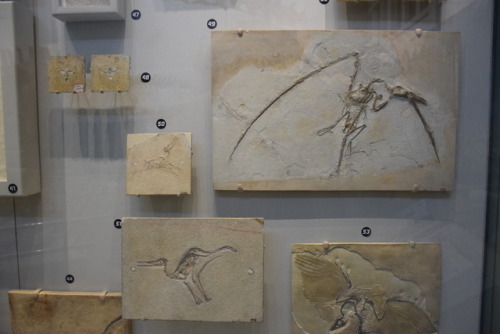
Three views of the most famous (and probably the most beautiful) Aspidorhynchus vs. Rhamphorhynchus fossil from the Upper Jurassic Solnhofen Limestone of southern Germany. Avid fisherman Matt Lamanna, the head of Vertebrate Paleontology at Carnegie Museum of Natural History (CMNH), jokes that the Aspidorhynchus looks angry, as if it’s mad about getting its snout stuck in the Rhamphorhynchus and dooming them both. Sorry Matt, this is just a quirk of preservation – the compression of the Aspidorhynchus skull during fossilization gave it the appearance of having grouchy eyebrows that weren’t there in life. You can learn more about this specimen in a paper by Frey and Tischlinger (2012).And if you want to see real fossils of both of these animals in person (albeit preserved separately), come visit the Solnhofen case in CMNH’s Dinosaurs in Their Time exhibition.

Because Aspidorhynchus lived only during the Mesozoic, there’s no chance that a modern-day angler will ever hook one. But should you find yourself fishing in one of Pennsylvania’s rivers or lakes this summer, and manage to land a gar or bowfin, pause for a moment and reflect on the ancient legacy of these fishes – a heritage that dates to the Age of Dinosaurs.

Lindsay Kastroll is a volunteer and paleontology student working in the Section of Vertebrate Paleontology at Carnegie Museum of Natural History. Museum staff, volunteers, and interns are encouraged to blog about their unique experiences and knowledge gained from working at the museum.

Related Content

Preserving Fossil Treasures: Eocene Fishes from Monte Bolca, Italy

Science Chats: Herpetologist Talks About Dinosaurs with Paleontologist

The Bromacker Fossil Project Part VIII: Martensius bromackerensis Honoring a Colleague

Filed Under: Blog Tagged With: dinosaurs in their time, Lindsay Kastroll, Mesozoic Monthly, Museum from Home, Science News, Vertebrate Paleontology

July 30, 2020 by wpengine

The Bromacker Fossil Project Part VIII: Martensius bromackerensis, Honoring a colleague

New to this series? Read The Bromacker Fossil Project Part I, Part II, Part III, Part IV, Part V, Part VI, and Part VII. 

Adult, holotype specimen of Martensius bromackerensis. Image digitally assembled by the author from five photographs taken by Diane Scott (Preparator at University of Toronto Mississauga [UTM]), 2010–2013. The specimen was collected in several large blocks.

The formal publication of some of the Bromacker discoveries took more time to complete than others, and our most recently pubished fossil, Martensius bromackerensis, holds the record in that regard. Four nearly complete specimens of Martensius were collected from the Bromacker quarry between 1995–2006. The first, discovered by Thomas Martens and his father Max, came from a jumbled pocket of fossils. Unfortunately, muddy groundwater had penetrated cracks in the subsurface of this portion of the quarry and coated and eroded bone present along these cracks. Despite this damage and the lack of a skull, we could identify the specimen as a caseid synapsid (synapsids, also known as mammal-like reptiles, are a group of amniotes whose later-occurring members gave rise to mammals).

Drawing of 1995 Martensius bromackerensis specimen. Because the specimen was collected in numerous pieces of rock, with parts of some bones exposed on apposing rocks, Scientific Illustrator Kevin Dupuis (UTM) had to first draw the bones exposed on each piece and then assemble all of the drawings digitally. Dotted lines indicate bone impression in the rock. Arrows point to healing scars from two fractures in the last right rib. Additional healing scars can be seen in preceding ribs. This animal apparently survived a serious injury. Modified from Berman et al., 2020.

The next specimen was discovered in 1999 by Georg Sommers (Preparator, Museum der Natur, Gotha), who prepared the fossil. It consists of a vertebral column, ribs, some limb bones, and a few scattered skull elements. Unfortunately, a more complete skull was needed to allow for comparison to other caseids, some of which are based only on skull material. It wasn’t until the discovery of two more specimens in 2004 and 2006 by Stuart Sumida and Dave Berman, respectively, that the long sought-after skull was found. Preparation of these specimens took a long time due to their size and the considerable amount of rock covering the bones in some of the blocks. My promotion to Collection Manager in 2005 left me with considerably less time to prepare fossils. Other preparators were asked to help with the preparation at both Carnegie Museum of Natural History (CMNH, Dan Pickering and Tyler Schlotterbeck) and in Dr. Robert Reisz’s lab at the University of Toronto at Mississauga (Diane Scott and Nicola Wong Ken). Robert was originally slated to lead the study, but other commitments prevented him from working on it, so Dave took over.

Besides preparation, the scientific study and publication of the specimens required illustrations and photographs, most of which were done by Diane, Nicola, and Kevin. Andrew McAfee (Scientific Illustrator, CMNH) made skeletal and flesh reconstructions of the animal, as well as an illustration of two Martensius in their ancient habitat (see The Bromacker Fossil Project Part III for a link to this illustration). All of this effort was worth it, however, because besides adding to the diversity of the Bromacker vertebrate fauna, Martensius has an unusual life history.

Juvenile specimen of Martensius bromackerensis. Image digitally assembled by the author from two photographs (skull and body) taken by Diane Scott in 2013. The skull, shown in ventral aspect, is incomplete and eroded on its dorsal surface.

Caseid synapsids are a diverse, long-lived group known from the Late Pennsylvanian–Middle Permian epochs (~300–259 million years ago) of Europe, Russia, and the USA, and, with one exception, all are adapted to eating plants (herbivorous). The most advanced caseids (such as the enormous Cotylorhynchus romeri) have ridiculously small skulls when compared to those of carnivores, spatulate (spoon-shaped) teeth tipped with small tubercles (cuspules) for cropping vegetation, and huge, barrel-shaped ribcages to support a large gut for fermenting cellulose-rich plants. The exception is the earliest known (Late Pennsylvanian epoch, ~300 million years ago) caseid, Eocasea martini, represented by a single, incomplete juvenile specimen from Kansas. The teeth of Eocasea are small and conical, which indicate that it most likely ate insects. Because it’s skull and ribcage are of normal size, in contrast to juveniles of Martenius, Eocasea probably ate insects throughout its life.

Reconstruction of the skull of Martensius bromackerensis (left) from the Early Permian (~290 million years ago) Bromacker quarry, Germany, and the more advanced caseid Ennatosaurus tecton (skull, middle and skull fragment with cuspule-tipped teeth, right), from the Middle Permian (~263 million years ago) of Russia. Skull reconstruction of Martensius made by Diane Scott and modified from Berman et al., 2020. Ennatosaurus skull reconstruction and jaw fragment drawing modified from Maddin et al., 2008.

Martensius has a modestly expanded ribcage and a small skull, suggesting that it was herbivorous. Furthermore, the feet of Martensius, like those of other caseids in which the feet are known, are large, with massive, elongated, strongly recurved claws. Martensius also has a well-supported hip region that may have enabled it to rise on its hind legs to reach and tear down overhead branches to feed upon.

The upper and lower teeth of the adult Martensius differ from those of more advanced caseids in being triangular and lacking cuspules. The upper jaw teeth of the juvenile resemble those of the adult, but the lower jaw teeth are more numerous—31 in the juvenile compared to 25 in the adult—and surprisingly, they resemble those of Eocasea. Dave concluded that juveniles of Martensius had teeth adapted for eating insects, which were replaced by an adult dentition that would’ve been good for cropping plants and piercing insects. Remarkably, the juvenile Martensius apparently died while in the process of replacing its juvenile dentition with that of adults.

So why have different juvenile and adult dentitions? Modern animals that eat fibrous plant matter have micro-organisms called fermentative endosymbionts in their large guts, which break down difficult-to-digest plant matter via fermentation. It is assumed that early fossil plant-eaters with broad ribcages also had large guts housing fermentative endosymbionts. Prior to the discovery of Martensius, other scientists hypothesized that early herbivores acquired endosymbionts by eating herbivorous insects that already had these microbes in their guts. In Martensius, the introduction of endosymbionts apparently occurred during the juvenile, insectivorous stage of life, which set the stage for adults to add plants to their diet.

Flesh (top) and skeletal (bottom) reconstructions of Martensius bromackerensis. Illustrations by Andrew McAfee and modified from Berman et al., 2020.

The generic name Martensius honors Thomas Martens for his discovery of vertebrate fossils at the Bromacker quarry and his perseverance in maintaining a highly successful, long-term field operation resulting in the discovery and publication of the exceptionally preserved Bromacker fossils. Bromackerensis refers to the Bromacker quarry, the only locality from which this species is known.

Stay tuned for my next post, which will feature some terrestrial dissorophoid amphibians.

For those of you who would like to learn more about Martensius, here’s a link to the 2020 Annals of Carnegie Museum publication in which it was described.

Amy Henrici is Collection Manager in the Section of Vertebrate Paleontology at Carnegie Museum of Natural History. Museum employees are encouraged to blog about their unique experiences and knowledge gained from working at the museum.

Keep Reading

The Bromacker Fossil Project Part IX: The Dissorophoid Amphibians Tambachia, Rotaryus, and Georgenthalia, Capable Travelers

Filed Under: Blog Tagged With: Amy Henrici, dinosaurs in their time, fossils, Museum from Home, paleontology, Science News, Section of Vertebrate Paleontology, Vertebrate Paleontology

July 15, 2020 by wpengine

The Bromacker Fossil Project Part VII: Eudibamus cursoris, the Original Two-legged Runner

New to this series? Read The Bromacker Fossil Project Part I, Part II, Part III, Part IV, Part V, and Part VI. 
Holotype specimen of Eudibamus cursoris, the most complete bolosaurid reptile known. Photo by the author, 2013.

Stuart Sumida discovered some small bones in the Bromacker quarry in 1993, the same year that the holotype skeleton of Diadectes absitus was found. Dave Berman told me that when Stuart showed them to him, he couldn’t see anything because they were so small. Upon closer examination, Dave, Stuart, and Thomas Martens identified them as those of the captorhinomorph reptile Thuringothyris mahlendorffae. Thomas’ wife Stefani, whose maiden name is Mahlendorff, discovered the first specimen in the Bromacker in 1982, and Thomas and a colleague named it in her honor in a 1991 publication.

The fossil was exposed in several pieces of rock, which Thomas shipped to Carnegie Museum of Natural History (CMNH) along with the large block of rock containing Diadectes. I didn’t prepare the specimen until several years later, as other projects, including the Diadectes, overshadowed it. Once I began working on it, though, Dave and I realized that it was not Thuringothyris. Indeed, we had no idea what type of animal it was, and our puzzlement grew as I exposed more of it. The identity wasn’t revealed until I had uncovered some very unusual, tiny teeth, which under the high magnification of the preparation microscope appeared to have a bulbous cusp towering over a basin. They looked vaguely familiar to me, but because I couldn’t immediately put a name on them, I rushed to get Dave from his office. Once Dave saw the teeth, he realized that the specimen was a new genus and species in the rare, enigmatic reptile group Bolosauridae.

Tiny teeth of a bolosaurid reptile, Bolosaurus striatus, in lateral (side; left) and occlusal (chewing surface; right) views. The specimen is in the CMNH Vertebrate Paleontology collection. Photos by Spencer Lucas (Research Associate, CMNH).

Until the discovery of Eudibamus cursoris, bolosaurids were represented in the fossil record by two genera, Bolosaurus and Belebey, which were based mainly on poorly preserved skull and fragmentary jaw fossils from Texas and Russia, respectively. Even though bolosaurids had been known since 1878, their relationship to other reptiles was not well understood. The nearly complete anatomy of Eudibamus allowed our team to determine that bolosaurids are the oldest member of the ancient group of reptiles called Parareptilia. This group has no living relatives, except possibly for turtles, a hypothesis that is highly debated by scientists.

Eudibamus cursoris fossil
Closeup of front and hind legs of Eudibamus. The hind leg, folded upon itself, is considerably longer than the front leg. Photo by the author, 2013.

When our study of the fossil began, we realized that Eudibamus was very different than other reptiles from that time. Proportions of the limbs and positions of the articulation surfaces on the upper and lower hind leg bones indicated that, in terms of posture, Eudibamus resembled a bow-legged human with a bad back instead of a typical sprawling reptile on four legs. It could stand and locomote on its hind legs in an upright posture (bipedal) with its legs held close together and in the same plane (parasagittal).

Dave was in constant phone communication with team member Dr. Robert Reisz (Professor, University of Toronto at Mississauga). One day Robert called Dave to ask if all the tail had been exposed, because he learned that modern lizards that are able to run bipedally have a long tail to help maintain their balance. The specimen was in Dave’s office and he immediately uncovered more of the tail and then let me finish the task. The tail was indeed very long and extended close to the edge of the block, which I had previously reduced in size. Additionally, we determined that the third, fourth, and fifth toes of the hind foot also were greatly elongated through lengthening of some of the individual toe bones, and that the first and second toes were extremely shortened by the reduction in size of individual toe bones. We hypothesized that when Eudibamus ran bipedally, it would rise on its toes, so that only the tips of the third, fourth, and fifth toes would contact the ground.

Drawing of the hind leg of Eudibamus cursoris (left) and the roughly contemporaneous reptile Captorhinus (right). Leg drawings are scaled to the same torso length of the whole animal. Illustrations of the animals are not to scale. Hind leg drawings are modified from Berman et al., 2000 and animal illustrations are from Wikimedia Commons.

Eudibamus occurred at least 60 million years before other bipedal, parasagittally-running reptiles appeared in the fossil record. This is reflected in its scientific name, which is derived from the Greek “eu,” meaning original or primitive, and “dibamos,” meaning on two legs. “Cursoris” is Latin, meaning runner. Examples of other reptiles using this locomotion mode are the dinosaurs Allosaurus fragilis and Tyrannosaurus rex, which you can view in CMNH’s Dinosaurs in Their Time exhibition.

So, what was the advantage of being able to run bipedally instead of running on all four legs? Lengthening the hind leg and foot would greatly increase stride length, especially if only the tips of the toes contacted the ground, which is an efficient way to increase speed. Eliminating arm to ground contact while running removes forelimbs from the path of the long-striding hind legs. The bulbous teeth and jaw structure of Eudibamusindicate that it was herbivorous. It seems likely, then, that Eudibamus used its ability to sprint to avoid becoming a tasty meal for a pursuing predator.

Eudibamus cursoris illustration
Peter Mildner (exhibit preparator at the Museum der Natur, Gotha) made a surprise visit to the Bromacker one afternoon to show us a model of Eudibamus cursoris he’d made. This image shows the model in the present day Bromacker quarry, part of the region it inhabited 290 million years ago. Photo by the author, 2006.

One of our laments is that a fossil trackway preserving Eudibamus walking quadrupedally and then switching to a bipedal gait has yet to be found.

Next time you are at CMNH, make sure you see the cast of the fossil skeleton and a model of Eudibamus that are exhibited in the Fossil Frontiers display case in CMNH’s Dinosaurs in Their Time exhibition. Stay tuned for my next post, which will feature the herbivorous mammal-like reptile Martensius bromackerensis.

For those of you who would like to learn more about Eudibamus, here is a link to the 2000 Science publication in which it was described: https://science.sciencemag.org/content/290/5493/969.

Amy Henrici is Collection Manager in the Section of Vertebrate Paleontology at Carnegie Museum of Natural History. Museum employees are encouraged to blog about their unique experiences and knowledge gained from working at the museum.

Keep Reading

The Bromacker Fossil Project Part VIII: Martensius bromackerensis, Honoring a Colleague

Filed Under: Blog Tagged With: Amy Henrici, dinosaurs in their time, Museum from Home, Science News, The Bromacker Fossil Project, Vertebrate Paleontology

June 29, 2020 by wpengine

Mesozoic Monthly: Protostega

June 20th was the first day of summer! The weather here in Pittsburgh is already beautiful. It’s enough to make one dream of a socially distant beach! Summer, of course, is sea turtle nesting season: during the next several weeks, female sea turtles all across our planet’s Northern Hemisphere will return to the beach where they hatched, drag themselves onto land, and lay their eggs in the sand. It would have been an incredible sight to see Protostega gigas, one of the largest sea turtles of all time, hauling itself onto the beach to lay its eggs! For June’s Mesozoic Monthly, we’re going to “dive in” to the paleontology of this giant reptile.

Carnegie Museum of Natural History’s spectacular skeleton of Protostega gigas is a composite made from the fossilized bones of two different individuals. Come see it on display in our Dinosaurs in Their Time exhibition when the museum reopens at the end of this month. But don’t forget to purchase your timed ticket in advance!

All turtles, including sea turtles like Protostega and tortoises like the Galápagos giant tortoise, belong to the group Testudines. This group originated during the Triassic Period, the first of the three time periods of the Mesozoic Era (aka the Age of Dinosaurs). Turtles split from other reptiles to form their own group before crocodiles and dinosaurs evolved! This means that turtles are not descended from dinosaurs, no matter how primordial some tortoises may look. Turtles differ from other reptiles in many ways, the most noticeable being their iconic shells. 

A turtle shell is formed of two main parts: the carapace, or top shell, and the plastron, or bottom shell. The shell is made of bone fused directly to the spine and ribcage, so a turtle cannot crawl out of its shell without leaving its skeleton behind! Another major difference between turtles and other modern reptiles involves skull anatomy. Turtles have anapsid skulls: the bony case that protects their brain lacks any external openings behind their eyes (known as temporal openings). All other extant reptiles plus birds are diapsids, meaning their skulls have two holes behind their eyes. Mammals differ from both conditions because we have only one temporal opening, making us synapsids. Traditionally, the anapsid condition of turtle skulls has been taken to indicate that they are the most primitive of living reptiles. More recently, however, many paleontologists and biologists have uncovered evidence that turtles are in fact diapsids whose evolutionary course led, for some reason, to a secondary closure of their temporal openings. According to these scientists, the closest relatives of turtles among today’s diapsids are either lepidosaurs (lizards, snakes, and kin) or archosaurs (crocodilians and birds).

A bird’s (or pterosaur’s!) eye view of Protostega gigas (left) swimming past two long-necked elasmosaurid plesiosaurs in shallow waters of North America’s Western Interior Seaway roughly 85 million years ago. (This scene is set in what’s now Kansas!) Art by Julio Lacerda; see more of his beautiful work here.

Reptiles, mammals, and birds all belong to a group called Amniota, and the key defining feature of amniotes is a protective layer around their eggs that allows this vulnerable life stage to survive on land. Having eggs that did not have to be laid in water meant that animals could move to less-wet habitats, a significant step in evolution! Unfortunately for sea turtles, which spend most of their lives at sea, this means they must return to land to lay their eggs. An amniotic egg would “drown” in water because the embryo still needs access to air. As a sea turtle, Protostega would have faced these same reproductive challenges, plus one more: it was huge!The largest modern turtle, the leatherback sea turtle, can grow over seven feet (2.1 meters) long; Protostega dwarfs it at 9.8 feet (3 meters)! If you’ve ever seen video of a sea turtle crawling onto the beach to nest, you know that it’s an awkward process. Imagine seeing a turtle that weighs at least a ton try to do the same! Although surely clumsy on land, Protostega was a graceful swimmer, using its four rigid flippers like wings to “fly” through the water.

Protostega lived in the Western Interior Seaway, an inland sea that stretched across much of North America during the Cretaceous Period (the third and final period of the Mesozoic Era). The seaway was warm, shallow, and teeming with all kinds of aquatic life: the perfect habitat for an omnivorous sea turtle. Because sea turtles are ectothermic (sometimes erroneously called “cold-blooded”), they cannot regulate their own body temperature. Instead, Protostega relied on warm water temperatures and sunlight hitting its back to keep warm. Although we don’t have a fossil record of the coloration of Protostega, we know that today’s large sea turtles are counter-shaded, with heat-absorbing, dark-colored backs and pale undersides. In an ocean environment where both predator and prey shift positions in the water column, this combination aids concealment. From below, a light-colored underside blends with light-saturated water. From above, a dark back blends with dark water. Camouflage in the water was an important feature when living alongside so many sizable predators. Protostega fossils have been found with bite marks from the large shark Cretoxyrhina mantelli, and it almost certainly was also on the menu for the mighty mosasaurs as well. Fortunately for us, we humans can enjoy the ocean knowing that few creatures are interested in eating us!

Lindsay Kastroll is a volunteer and paleontology student working in the Section of Vertebrate Paleontology at Carnegie Museum of Natural History. Museum staff, volunteers, and interns are encouraged to blog about their unique experiences and knowledge gained from working at the museum.

Related Content

Mesozoic Monthly: Citipati

Science Chats: Herpetologist Talks Dinosaurs with Paleontologist 

The Volunteers: Can’t Live Without Them

Filed Under: Blog Tagged With: Cretaceous Seaway, dinosaurs in their time, fossils, Lindsay Kastroll, Museum from Home, Science News, Section of Vertebrate Paleontology

May 27, 2020 by wpengine

Mesozoic Monthly: Citipati

The month of May that we’re living in is very different from the one we all anticipated at the start of the year. However, society somehow manages to march on. College students are still graduating, moms are still being celebrated, and Mesozoic Monthly continues! Our honoree for the month of May is known to have been a dedicated parent due to several specimens that show adults guarding eggs. Say hello to Citipati osmolskae!

illustration of Citipati, a dinosaur that looks similar to a bird, on a nest of blue eggs

A devoted Citipati parent guarding its nest. Some evidence suggests that the Citipati skeletons found atop nests may have been males (rather than females as was originally thought). Also, recent research indicates that—believe it or not—oviraptorid eggs were blue! Art by ginjaraptor on DeviantArt.

It might not look like it, but Citipati is a theropod, like the more famous dinosaurs Tyrannosaurus, Allosaurus, and Velociraptor. Most theropods were carnivores, sporting skulls with big toothy grins, but not all theropods were ravenous predators! There are several groups of theropods that evolved toothless beaks for specialized diets. One of the stars of Jurassic Park, Gallimimus, was part of a predominantly herbivorous group of beaked theropods called Ornithomimidae. Citipati belongs to another group of beaked theropods called Oviraptoridae. “Oviraptor” means “egg thief,” in reference to an old hypothesis that oviraptorids stole and ate eggs from other dinosaurs’ nests. The discovery of a Citipati skeleton perched in a brooding position atop a nest of eggs was pivotal in changing this idea. We now know that instead of stealing others’ eggs to eat, fossilized oviraptorids preserved near eggs were actually protecting their own eggs! The eggs in an oviraptorid’s nest were arranged in circles with a space in the center for the parent to sit and spread their feathered arms over their incubating young.

photograph of Anzu dinosaur fossil

Citipati and other oviraptorids are closely related to one of Carnegie Museum of Natural History’s most bizarre dinosaurs, the ‘Chicken from Hell’ Anzu wyliei, shown here on display in the museum’s Dinosaurs in Their Time exhibition.

So, instead of eggs, what would the toothless beak of Citipati have been used to eat? Because most oviraptorid beaks are very deep, like those of modern parrots, most paleontologists infer that these dinosaurs ate mostly plants. However, this doesn’t necessarily mean that meat was off the menu; it would still have been possible for oviraptorids to have eaten small animals, making them omnivores. On top of its thick skull, Citipati possessed a tall, triangular crest that gave its small head a square-shaped profile. This crest was not as impressive as those on some other dinosaurs, but since Citipati grew to ten feet (three meters) long, the animal would still have been quite imposing. I certainly wouldn’t want to get between a Citipati parent and its eggs!

Citipati fossils are found in the modern Gobi Desert of Mongolia, in rocks known as the Djadokhta Formation. The Djadokhta rocks are made of sediments that were deposited late in the Cretaceous Period, preserving details of the ecosystem that existed there roughly 80–75 million years ago. The name Citipati means “funeral pyre lord,” which is fitting due to the hot environment in which this oviraptorid lived. Also, Citipati shares its name with a Buddhist deity that is believed to protect cemeteries from thieves, which is an appropriate parallel considering how the skeleton of this dinosaur was found guarding its fossilized nest.

Although the habitat Citipati lived in was a desert, like the Gobi Desert that is there today, this prehistoric desert was probably not as dry. In the event of rain, water gathered in temporary streams that drained the water to basins and oases. Since desert rain events are by definition few and far between, any animals that did not live near these oases would have needed to have adaptations for going without water for a long period of time. Some of the animals that lived in this unwelcoming environment alongside Citipati included everyone’s favorite small theropod Velociraptor, the hornless ceratopsian Protoceratops, and the tail-club wielding ankylosaur Pinacosaurus.

Lindsay Kastroll is a volunteer and paleontology student working in the Section of Vertebrate Paleontology at Carnegie Museum of Natural History. Museum staff, volunteers, and interns are encouraged to blog about their unique experiences and knowledge gained from working at the museum.

Filed Under: Blog Tagged With: dinosaurs in their time, Lindsay Kastroll, Mesozoic Monthly, Museum from Home, Science News, Vertebrate Paleontology

May 14, 2020 by wpengine

The Bromacker Fossil Project Part II: The Hunt for Fossils

New to this series? Read The Bromacker Fossil Project Part 1 here.

Finding fossils at the Bromacker quarry was tedious and physically demanding, but it was extremely rewarding when a fossil is discovered. Our annual summer field season generally lasted three and a half weeks. Because the weather usually wasn’t conducive to camping or cooking outdoors, we stayed at the same hotel and dined at the hotel or local restaurants.

The original 1993 fossil quarry was opened using heavy equipment and operators from the nearby commercial quarry, and in the early years we relied on these people to expand the fossil quarry’s boundaries as needed. When the commercial quarry was temporarily shut down due to the lack of contracts for building stone, our collaborator Dr. Thomas Martens fortunately was able to obtain funding to annually rent a Bobcat, which he became skilled at operating. Thereafter, Thomas would use the Bobcat to expand the quarry and remove soil and weathered rock layers, so that we could begin our yearly excavation on unweathered rock.

image
The Bromacker quarry on the first day of fieldwork in the 2006 field season. Shovels were used to clear loose rock from the surface of the quarry. Pictured (counterclockwise from left) graduate student Andrej Čerňanský and Dr. Jozef Klembara (Comenius University, Bratislava, Slovak Republic) and Dr. Dave Berman (Carnegie Museum of Natural History [CMNH]). Photo by the author, 2006.

We would each stake out an area of the quarry to work in and then proceed to work through the rock layers by using a hammer and chisel or pry bar to free a piece of rock. Its surfaces and edges would be checked for fossil bone, and if there was none, the rock piece would be broken into smaller pieces, which were also checked for bone. As is the case at many other fossil sites, the rock tended to split along the plane a fossil was preserved in, because the fossil would create a zone of weakness.

image
The quarry after a couple days of excavation. Pictured (clockwise from front) are Dave Berman, Jozef Klembara, and Andrej Čerňanský. Photo by the author, 2006.

Once a fossil specimen was discovered—and there were a few frustrating years when this didn’t happen—the hard work of extracting it from the quarry began. Here, I’ll use a fossil discovered during the 2006 field season as an example of how this was done.

image
A discovery! Fossil bone and bone impression are exposed to the left of the lens cap. Photo by the author, 2006.

First, we would isolate the fossil specimen from the surrounding rock, exposing as little of the fossil as possible while determining its extent, because it would have been easy to lose pieces of bone in the dirt and mud. Then we would encase the specimen in a plaster and burlap jacket to protect it during extraction, shipping, and preparation.

To make the jacket, we’d coat cut strips of burlap in wet plaster and then spread them across the surfaces of the rock containing the specimen, or block. A layer of plastic (plastic bags worked well) was applied to the top surface of the block to keep plaster from sticking to any exposed bone.

image
The block is partially isolated from surrounding rock. In this case, we decided to encase the top and some sides of the block in plaster and burlap bandages to hold the rock pieces together before we finished isolating the block from surrounding rock. Photo by the author, 2006.

After a couple layers of plaster bandages were applied to the top and sides of the block, the block was undercut, with plaster bandages added periodically to hold the undercut rock in place.

image
Pictured (left to right) are Dr. Stuart Sumida (California State University, San Bernardino, CA), Dave Berman, and Mr. Jerome Gores (Museum der Natur, Gotha [MNG]). Jerome is holding a plaster and burlap bandage while Dave and Stuart are pressing plaster bandages against the bottom of the block. They must hold the bandages in place until the plaster sets. Photo by the author, 2006.
image
Using hammers and chisels to undercut the block. Photo by the author, 2006.

When deemed safe, we would crack the block free from the quarry floor using hammers and chisels, and flip it over, unless it broke free on its own. Excess rock would be removed from the bottom of the block to make it lighter in weight. Then we would apply burlap and plaster bandages to the bottom of the block. The block would be removed from the quarry and stored at the MNG until it was shipped to Pittsburgh.

image
The block has now been safely flipped over and excess rock is being removed. Photo by the author, 2006.

We encountered several problems during our quarry operations over the years. As we worked our way through the rock column in the quarry, processed rock piled up on the quarry floor. In the early years, we tossed or shoveled the processed rock into wheelbarrows and pushed the heavy, unwieldy wheelbarrows out of the quarry to a dump pile. Fortunately, the Bobcat eventually replaced the wheelbarrows for moving processed rock. As we ran out of space outside the quarry to dump processed rock, the rock was used to backfill older portions of the quarry.

image
Former CMNH volunteer Linda Rickets (front, right) and the author (left, rear) line up to push loaded wheelbarrows out of the quarry to the dump pile. Photo by Dave Berman, 1996.
image
At the dump pile. Over time rain and the freeze/thaw cycle would break down the rock and vegetation would grow on it. Photo by Dave Berman, 1996.

Another problem was that fossils found at the bottom of the quarry were often extremely difficult to undercut because the rock so was hard. Sometimes a well-hit chisel would just bounce off the rock instead of cracking or penetrating it. One year we had to resort to a rock saw to undercut a block.

image
Dave Berman uses a rock saw to undercut a block. Photo by the author, 2004.

Rain was always a problem. We would shelter in our cars during intervals of rain, or work at the museum if the rain was heavy and persistent. Occasionally heavy rain would flood the quarry, forcing us to work in the ‘dry’ areas of the quarry while a pump drained the water. Of course, we had contests to see who could skip a rock the farthest or make the biggest splash.

image
A flooded quarry greeted us in the morning after heavy overnight rain. Pictured are Thomas Martens (front) and Stuart Sumida (rear). Photo by the author, 2010.

Next week’s post will describe the process of fossil preparation, that is, removing rock to reveal a specimen in the lab. The fossil collected in the 2006 field season will be used as an example.

Here are some videos taken by taken by Thomas Martens’ wife, Steffi, during the 1993 and 2006 field seasons. These show the process of searching for fossils (1993 video) and collecting the fossil highlighted in this post (2006 videos).

Bromacker Quarry 1993

https://youtu.be/DAEG0l1NotE

Bromacker Quarry 2006

https://youtu.be/UNL1s5ycJSM

https://youtu.be/lqYxheOZLQY

https://youtu.be/HrmSBrhde_E

Amy Henrici is Collection Manager in the Section of Vertebrate Paleontology at Carnegie Museum of Natural History. Museum staff, volunteers, and interns are encouraged to blog about their unique experiences and knowledge gained from working at the museum.

Keep Reading

The Bromacker Fossil Project Part III: Fossil Preparation

Filed Under: Blog Tagged With: Amy Henrici, dinosaurs in their time, fossils, Museum from Home, Science News, Vertebrate Paleontology

  • « Go to Previous Page
  • Page 1
  • Interim pages omitted …
  • Page 6
  • Page 7
  • Page 8
  • Page 9
  • Page 10
  • Interim pages omitted …
  • Page 15
  • Go to Next Page »

sidebar

About

  • Mission & Commitments
  • Directors Team
  • Museum History

Get Involved

  • Volunteer
  • Membership
  • Carnegie Discoverers
  • Donate
  • Employment
  • Events

Bring a Group

  • Groups of 10 or More
  • Birthday Parties at the Museum
  • Field Trips

Powdermill

  • Powdermill Nature Reserve
  • Powdermill Field Trips
  • Powdermill Staff
  • Research at Powdermill

More Information

  • Image Permission Requests
  • Science Stories
  • Accessibility
  • Shopping Cart
  • Contact
  • Visitor Policies
Stay in the loop! Sign up for our newsletter(s).
One of the Four Carnegie Museums | © Carnegie Institute | Privacy Policy | Terms of Use | Accessibility
Rad works here logo