• Skip to primary navigation
  • Skip to main content
  • Skip to primary sidebar

Carnegie Museum of Natural History

One of the Four Carnegie Museums of Pittsburgh

  • Visit
    • Buy Tickets
    • Visitor Information
    • Exhibitions
    • Events
    • Dining at the Museum
    • Celebrate at the Museum
    • Powdermill Nature Reserve
    • Event Venue Rental
  • Learn
    • Field Trips
    • Educator Information
    • Programs at the Museum
    • Bring the Museum to You
    • Guided Programs FAQ
    • Programs Online
    • Climate and Rural Systems Partnership
  • Research
    • Scientific Sections
    • Science Stories
    • Science Videos
    • Senior Science & Research Staff
    • Museum Library
    • Science Seminars
    • Scientific Publications
    • Specimen and Artifact Identification
  • About
    • Mission & Commitments
    • Directors Team
    • Museum History
  • Tickets
  • Give
  • Shop

Super Science Days

October 13, 2020 by wpengine

Invasion of the Leaf Galls!

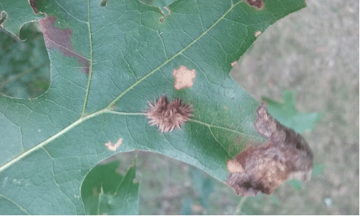
Happy Gall-oween! Mwah-hahaha! Prepare yourselves for the silent invasion of the leaf galls! Over the summer and into the early fall, you may have seen something very strange happening to the oak trees of Southwestern Pennsylvania. Small, furry growths, brown or orange in color, have been appearing on oak leaves. If you haven’t seen them, imagine a Tribble from Star Trek, but in miniature size growing directly on the leaves of a shady oak.

These are leaf galls—but they aren’t an alien lifeform nor a devastating tree blight. They are the product of a fascinating chemical reaction.

Early in the spring, just as the oak trees are beginning to bud, gall wasps (from the family Cynipidae) lay their eggs on the brand-new leaves. These creatures—smaller than a fruit fly and lacking the ability to sting—might also lay their eggs on the twigs of the trees or on the stems of goldenrod. Once the wasp eggs hatch, the larvae begin to eat the leaf on which they were deposited. This is when things get interesting: when the chemicals in the larvae’s saliva mingle with the plant hormones in the leaf, the gall begins to form. Depending on the drop site and the species of gall wasp (there are over 700 species in the United States alone that target oaks), the appearance of the gall will be different. For instance, when the eggs hatch on a branch or twig and begin their feast, the gall will have a dense, spherical appearance. This is the specific kind of gall that gives the phenomenon its name: “galla” means “oak-apple” in Latin.  Some leaf galls might take on the shape of tiny brown flying saucers as they did in Jefferson and Forest Counties in recent years. Some other galls have the appearance of spindly red fingers or peppers protruding from the leaf. The variety of tree and leaf galls are, in a word, kaleidoscopic.

While there is great variation in the physical appearance and structure of leaf galls, they each serve a shared purpose. The chemicals that the larvae secrete as they “chew” stimulate the leaf into creating a gall for shelter and sustenance. The gall is a protective, nutrient-providing dome over the developing larvae. While the galls sometimes interrupt the process of photosynthesis and cause some leaf browning and curling, they won’t kill the tree itself. The gall wasp is a mostly benign parasite. By mid-October, the wasp-bearing galls will fall from, or with, their leaves. The next spring, the surviving wasps will emerge from the soil.

leaf galls on green leaf

Some years, this new generation will breed sexually. Other years, it will be entirely female and reproduce asexually. That is, through parthenogenesis, the same process that the dinosaurs in the original Jurassic Park reproduce. Remember Dr. Malcolm’s famous “life finds a way” monologue? Galls are misunderstood by the general public because they perceive the phenomenon as a nuisance and eyesore. Scientists warn against treating infested trees with pesticide or scraping off the galls. Such actions would do more harm than good to the trees. Instead of being an unnerving menace, the gall wasp is an awe-inspiring example of how one animal uses its surrounding ecosystem—without excessive harm—to ensure that its kind will perpetuate itself safely and successfully.  Furthermore, the weirdly wonderful shapes and designs of the leaf gall demonstrate that nature isn’t just useful but also beautiful.  It’s that beauty that makes this seemingly bizarre invader more than a seasonal annoyance.

Nicholas Sauer is a Natural History Interpreter and Gallery Experiences Presenter at the Carnegie Museum of Natural History. Museum employees are encouraged to blog about their unique experiences and knowledge gained from working at the museum.

Related Content

Booseum: Spooky Coloring Pages

Booseum: Make Snail Slime

Scientist Takeover: Invertebrate Zoology Specimens

Filed Under: Blog Tagged With: booseum, Educator Resources, Educators, Nicholas Sauer, Super Science Days

September 18, 2020 by Kathleen

Scientist Takeover: Invertebrate Zoology Specimens

Murray Crayfish

Murray Crayfish

The Murray Crayfish (Euastacus armatus) is a favorite of Jim Fetzner, Assistant Curator of Invertebrate Zoology.  It can be found in the Murray and Murrumbidgee River catchments in the Australian states of New South Wales and Victoria.  The species was originally described back in 1866 by Eduard von Martens. The species is becoming rare due to habitat degradation and overfishing and is considered Threatened or Endangered by Australian conservation agencies. The species is one of the largest species of freshwater crayfish in the world, second only to the Giant Tasmainan Freshwater Crayfish, Astacopsis gouldi. It can reach maximum sizes of over 4 pounds and about 16 inches in length.  The claws are typically bright white, and the body is usually black or dark greenish/brown and covered in large spines, making it a quite striking crayfish when seen in the wild.

Lasiocampidae moths

Lasiocampidae moths

Lasiocampide is the favorite moth family of Vanessa Verdecia, Scientific Preparator in the Section of Invertebrate Zoology.  There are approximately 2,000 species worldwide which include moths commonly referred to as the eggars, lappet, and tent caterpillar moths.  Check out this drawer which includes mixed species that need to be curated into the main collection.  These moths have reduced mouthparts and do not feed as adults, so all the eating is done in the caterpillar stage.  Some species in this family are well known, including the Eastern Tent Caterpillar (tray 3 and 4), which is a pest.  However, there is interesting species diversity in the Tropics, with species waiting to be discovered and named.  Although there are only 35 species in the US, questions remain about the life cycles and number of species in some of the groups.  Field work and molecular data using specimens in the Carnegie collection will help to answer these questions and revise studies that have been published in the past.

Bold Jumping Spider

Bold Jumpers

Phidippus audax, commonly known as the Bold Jumper, is one of the favorite species of invertebrates of Catherine Giles, Curatorial Assistant in the Section of Invertebrate Zoology. They’re very common in North America, but what makes them stand out from others is their iridescent chelicerae—their “jaws.” Members of the genus Phidippus can all easily be identified by this iridescence, and typically males will have brighter iridescence to attract a mate. P. audax is very docile with some people even keeping them as pets! Spiders are always handy to have around as they eat problematic insects, like mosquitoes. Specimens of P. audax in our spider collection have been found in nearby Frick Park. See if you can spot these shiny-faced little ones on your next walk through the park!

Manticora adult beetle

Manticora, Adult Beetle

Manticora imperator is a tiger beetle in the family Carabidae, and a favorite of Bob Davidson, Collection Manager Emeritus in the Section of Invertebrate Zoology. The genus Manticora (“the one who devours men”) consists of 15 known species confined to the southern portions of Africa, mostly to the oldest geologic portions of that region, and mostly to open desert and dry savannah habitats. They are relatively primitive, flightless, predatory black tiger beetles of enormous size. The males of some species are particularly spectacular, with huge asymmetrical mandibles, reaching the extreme in Manticora imperator, with a toothed left mandible and a larger right mandible bent like a sickle. Mandibles in both sexes are used to attack prey, and, in males, also to combat other males and to clasp the female during copulation.

Manticora beetle larva

Manticora, Beetle Larva

The larva of Manticora mygaloides, one of 15 known species in this genus which is only known from the southern portions of Africa.  The Manticora larvae look and behave more like tiger beetle larvae from other parts of the world, except that they are enormous.  They mostly occur in open desert and dry savannah habitats, where they dig a vertical burrow up to a meter in depth, depending on substrate, which they can drop down into when disturbed.  The larval head is like a big armored plug with jaws attached.  In attack mode, they block the burrow entrance with the head (making the hole difficult to see) and wait.  There is also a large hook toward the rear on the larva’s back which makes it difficult for anything to dislodge it from the burrow. If something edible gets within striking distance, the larva throws its forebody out, grabs with its large jaws, and drags the prey into the burrow.

Deaths Head Hawkmoths

Death’s Head Hawkmoths

The African Death’s Head Hawmoths, Acherontia atropos, are found in Europe and Africa.  They are members of the family Sphingidae, which include about 1,450 species commonly referred to as the hawkmoths, sphinx moths, and hornworms.  There are two other species in this genus—A. lachesis and A. styx, which are found in Asia.  All three species are known for the skull-like color pattern formed by the scales on the thorax and the rib-like color patterns on the abdomen, which have inspired stories and superstitions in the regions of the world where they occur.  Acherontia styx was referenced in the book The Silence of the Lambs by Thomas Harris.  A scene of the movie adaptation was filmed in the Section of Invertebrate Zoology which depicted an entomologist identifying the pupa of a sphingid moth.  The drawer imaged here includes specimens in the caterpillar and pupal stages of Acherontia atropos, prepared and preserved dry according to historical standards.  Larval and pupal specimens are now preserved in ethanol.  All three species are known for their interesting biological adaptions including a mechanism that allows them to squeak, and the ability to feed on honey which they steal from the combs of honeybees.

Saturniidae moths

Saturniidae moths

The family Saturniidae, which includes about 2,300 known species, are commonly referred to as the royal, emperor, and giant silk moths.  They are known for their large size, colorful scale patterns, and some have “eyespots” on the hindwings that serve as a defense mechanism for scaring off predators.  The adults have reduced mouth parts, so they do all their feeding in the caterpillar stage and cannot feed as adults.  Therefore, they only live for a few days in the adult stage—long enough to mate.  Pictured here are some of the species known to occur in Pennsylvania.  The Pine Devil Moth (tray 1) and the Royal Walnut Moth (tray 3) are closely related and there is evidence of population decline, especially in the Northeastern United States.  They have horns that look scary but are harmless.  The Imperial Moth (tray 2) has also experienced population decline and has four color forms seen in the caterpillars.  One culture may include dark brown, light brown, red, and green caterpillars all from a single parent!  The Cecropia Moth (tray 4) is very common in Pennsylvania but has also experienced population decline that is thought to be due to parasitism by a tachinid fly introduced to control Gypsy Moths, which are an introduced pest that threatens our forests.

Pachyrhynchus specimens

Pachyrhynchus specimens

Members of the genus Pachyrhynchus are the favorites of Ainsley Seago, Associate Curator of Invertebrate Zoology (who calls them “disco weevils”).  The glittering colors of these party beetles come from tiny photonic crystals inside their flattened scales; several of Bob Androw’s cerambycids have independently evolved a similar structural color mechanism.  No one is sure what these Indonesian weevils use their colors for, but it may be a signal warning predators not to bother… their fused elytra and tough exoskeleton are too thick to pierce.

Sternotomus callais

Sternotomus callais

The longhorn beetle genus Sternotomis has convergently evolved structural colors based on three-dimensional photonic crystals, just like those of Pachyrrhynchus but arising from a different ancestral lineage. These colors are created by the nanoscale interactions of photons with a crystalline structure within the beetle’s flattened hairs (“setae”), and will last as long as the specimen itself. Paleontologists have even found fossil beetles that retain their iridescence after 60 million years!

Longhorn Beetles

Longhorn Beetles

The longhorn beetles (Cerambycidae) are a favorite of Bob Androw, Collection Manager in the Section of Invertebrate Zoology.  Although these beetles are typically known for their very long antennae, that character varies and quite a few species have short antennae. 

Check out this drawer of diverse looking specimens from the Carnegie collection which represents just a few of the 26,000 species known.  It’s a large taxonomically diverse and economically important family of beetles which contains many common and showy species, some serious plant pests, and a number of species that are considered rare enough to be afforded legal protection.

Some species mimic other insects, such as ants, bees, and wasps in both shape and coloration.  The larvae are mostly wood-boring and occur in dead or decaying wood, with a few species feeding on live plant tissue.  A few are soil-dwelling as larvae, feeding on the roots of grasses and other plants. The adult beetles have a wide range of feeding habits that include visiting flowers for nectar and pollen; feeding on fruits and sap from trees; and feeding on bark, stems, and leaves. There are also some that don’t feed at all as adults and just one genus, Elytroleptus, is known to be carnivorous as a predator on net-winged beetles (family Lycidae).

A: Moneilema sp. – Subfamily Lamiinae. These flightless species breed as larvae in the living tissue of cactus in the Southwest United States and Mexico. The adults are mimetic of darkling beetles like Eleodes spp., in the beetle family Tenebrionidae.

B: Enoplocerus armillatus – Subfamily Prioninae. This is one of the largest species of Cerambycidae in the New World, surpassed only by the enormous Titanus giganteus. Both occur throughout the Amazon Basin, while Enoplocerus ranges north as far as Costa Rica.

C: Callisphyris sp. – Subfamily Cerambycinae. A spectacular example of mimicry of a wasp by a cerambycid, involving coloration and the fuzzy hindlegs.

D: Hypocephalus armatus – Subfamily Anoplodermatinae. One of the most atypical species of cerambycids, possessing very short antennae, legs adapted for digging and an oddly shaped body. It is primarily soil-dwelling, occurring in the northern parts of South America.

E: Aphrodisium cantori – Subfamily Cerambycinae. While such brilliant metallic colors make this species stand out against a white background, it would well camouflaged in its natural habitat, sitting on a green leaf in a sun-dappled jungle in Southeast Asia.

F: Petrognatha gigas – Subfamily Lamiinae. Native to tropical Africa, this species can almost disappear while sitting on charred wood despite its size. Females are attracted to recently burned areas where they deposit eggs in the damaged wood as a host for their larvae.

G: Onychocerus scorpio – Subfamily Lamiinae. This South American species is a great  example of cryptic coloration which allows it to blend into its surroundings when sitting on dead wood.

H: Acrocinus longimanus – Subfamily Lamiinae. The spectacularly elongate forelegs of this species make it notable amongst the Cerambycidae. The evolutionary driver for this is unknown, and no behavior involving these long appendages differing from other cerambycids has been observed.

I: Rosalia alpina – Subfamily Cerambycinae. This beautiful blue species is found across the European continent but is restricted to old-growth forests, leading to a decline in numbers. It is protected by law in a number of countries. The closely related Rosalia funebris (L) is found in the western United States, and while not uncommon, is not often observed. Can you find the third species, Rosalia batesi, from Japan, in the drawer?

J: Acanthocinus aedilis – Subfamily Lamiinae. The source of the common name of “long-horned beetle” is obvious in this species. It has one of the longest antennae-to-body length ratios in the Cerambycidae.

K: Leptura quadrifasciata – Subfamily Lepturinae. This common species is an example of the lepturines, the “flower longhorns” – showing a common color pattern mimicking the pattern of bees or wasps. The subfamily contains many species that are diurnal pollen feeders.

Filed Under: Blog Tagged With: Scientist Takeover, Super Science Days

September 18, 2020 by Kathleen

Scientist Takeover Activity: DIY Field Kit

Field Work

Research can take on many forms and occur in a variety of locations. We can conduct research through controlled experiments in sterile labs, or by closely reading of historical texts found in archives, among other research methods. Research can also occur “in the field.” This type of research is often called fieldwork, field research, or field study. Fieldwork in natural sciences like biology, geology, or ecology is used to understand how natural environments function. It involves the observation and collection of data about organisms and habitats in their everyday settings. 

Fieldwork requires a good deal of preparation. Along with planning accommodations, food, clothing, medical supplies, and the other usual travel needs, researchers conducting fieldwork also have to prepare for their on-site scientific investigations. To do this, they bring all the necessary gear required for observing, recording, collecting, etc. Some of the standard equipment is more highly specialized and expensive like GPS and certain measuring devices. However, most of the tools used in field work are similar to items easily purchased at your local grocery or hobby store. This means that we can actually make low-cost versions of these fieldwork kits to use in our very own backyards.

gloves, measuring tape, camera, tape, jar, notebook and pens

DIY Field Kit

Observing

When you are out in the field, your main task is to detect or discover. You might want to look for a particular species or maybe a pattern of interactions. To do this, you can mostly rely on your own senses of sight, smell, touch, and hearing (taste might also be useful in some instances, but we advise against tasting anything without consulting a professional). However, it can be helpful to have some tools that enhance your observational senses.

  • Binoculars and/or Magnifying Glass – Though our eyes are useful tools, they sometimes need help seeing things outside our normal abilities during observation. Binoculars are helpful for seeing higher detail of things in the distance. Magnifying glasses help enlarge finer details on objects up close.
  • Naturalist Guide Book or App – Print and online guides for observing can aid in knowing what to look for, where to look, and identifying what you observe. There are numerous options available at your local library as well as retailers. 

Recording

After you have observed something new, exciting, or simply something you want to remember, you need to record information about your findings. This step is key for future comparison, developing plans for ongoing observation, and keeping track of the things you have already observed.

  • Pencils and Pens– Always have a few writing implements with you during field work. You would not want to be empty handed when you see something important!
  • Notepad or Notebook – Smaller sizes can be more convenient for storage and movement in the field. However, any size will do. You will want to record the date, location, and details about your observations in the field in these notebooks. These entries can include sketches, descriptions of sights/sounds/smells/etc., and any other details that you find important.
  • Ruler or Measuring Tape (optional) – If you want to be fairly precise in your notes, you might want to have a tool for measuring specimens.
  • Camera (optional) – A camera can be useful for documenting a scene or specimen in nature. If you plan to use a camera, be careful with any sudden or invasive movements that might disturb the scene you hope to capture.

Collecting

Collecting samples may be useful for continued examination.

  • Gloves – Unless you know exactly what you are collecting, it is always a good idea to protect yourself. Use gloves when interacting with your samples.
  • Scissors – Depending on what you collect, scissors may not be necessary.
  • Glass and/or Plastic Containers – You will need some place to store your samples once collected. Rigid, hard containers will better ensure the safety of your samples, but plastic bags can also be used.
  • Labeling Marker and Tape – Always make sure to label your samples with date and location information. This way, you can more easily identify and compare your samples with your notes and other samples.

And last, but not least is the most important tool in your field kit… Patience. Patience is not technically a tool you can purchase, but it is extremely important for field work. It can sometimes take days, weeks, or even years to observe certain phenomena.

Now, put all these items in a case of your choosing (one with sections for organization, if possible) and see what you can find! 

This activity was written by Jane Thaler, a Gallery Presenter in the museum’s LifeLong Learning Department.

Filed Under: Blog Tagged With: Scientist Takeover, Super Science Days

September 18, 2020 by wpengine

In the Field: Following the Work of a Paleontologist

Introduction by Jessica Sperdute

Edited by Matt Lamanna

With 22 million specimens housed at Carnegie Museum of Natural History (CMNH) and nearly 10,000 on display at any given time, chances are you’ve seen a dinosaur or two during your museum visits. But have you ever wondered how those dinosaurs get to the museum after they’re found? Or how we know where to dig for them in the first place?

What is a Fossil?

Fossils are the remains of animals, plants, and other ancient life that have been preserved in rock layers, or sediment. Fossils can include things such as leaves, skin, feathers, hair, footprints, and, most commonly, hard material such as wood, shells, teeth, and bones. Even poop can be fossilized! Many kinds of fossils are rare, and studying them can help us understand how the world looked tens of thousands or even millions of years before our time. Scientists who study fossils are known as paleontologists.

Looking at the Layers

Paleontologists use many tools to help them find fossils, but the key to knowing where fossils may be hidden underground lies with rocks—massive layers of rocks, called strata, are piled onto one another over time. These layers of different rocks can tell us not only what type of rock the layer is made of, but also approximately how old the layer is. The study of rock layers is called stratigraphy, and paleontologists use it to find potential fossil beds. For instance, if a paleontologist is looking specifically for fossils of dinosaurs, they would use stratigraphy to locate exposed layers of sedimentary rocks that formed at the time when dinosaurs lived and died—the Mesozoic Era. Once rocks from the Mesozoic Era are found in a location, the paleontologist goes to that location to hunt for fossils.

Big Prospects

Finding the right type of strata is only half the work of finding fossils; once paleontologists arrive at the field site, they need to physically walk around and search for clues that fossils may be around or underneath them. This is called prospecting, and the best place to prospect is usually at the base of a hill. Wind and rain will erode or gradually wear away rocks, allowing some fossils to break loose from higher sediments and roll downhill. If a fossil fragment is found, the team can then search the area to see if there may be other, more complete fossils—oftentimes higher up the hill and still embedded in rock.

Once prospecting has yielded an area where a fossil is likely to buried, the team can begin to block out the site and start digging. They use a wide variety of tools—even household items like paintbrushes, shovels, and hammers—to uncover fossils without damaging them. Records are taken of this step-by-step process to ensure all the data, from the precise location of the dig site, to the type of fossils found and their spatial relationships to one another, and even the measurements of the quarry, is kept for further study.

Safety First

The team has found a fossil, dug it up, and recorded the data. Now what? Once a fossil has been carefully excavated, it needs to be protected. Most fossils are delicate, so to transport them, especially larger ones, paleontologists use a method called plaster jacketing to protect them. First, they wrap the fossil in soft material such as paper towels, toilet paper, or aluminum foil to cover it. Then they wrap the covered fossil in strips of burlap that have been soaked in liquid plaster. This method is like using a cast on broken bones. After the plaster hardens, it acts as a shield. When the fossil has been safely transported and is ready to be studied or put on display at a place like Carnegie Museum, the paleontologist can gently cut away the plaster without damaging the fossil inside.

Paleontologist Photos

Dr. Matt Lamanna, Mary R. Dawson Associate Curator of Vertebrate Paleontology here at CMNH, has shared some of his favorite photos of his work at previous fossil dig sites. Look at the photos—do you recognize some of the locations, the tools that Dr. Lamanna is using, or the fossils that he’s digging up?

Here, Carnegie Museum of Natural History Mary R. Dawson Associate Curator of Vertebrate Paleontology Dr. Matt Lamanna is pointing at two ribs of a small—possibly baby—sauropod (long-necked plant-eating dinosaur) projecting from a rock face in the Bahariya Oasis of Egypt in 2001. He’d found this small sauropod only minutes before this photo was taken. Sometimes prospecting yields great finds! Credit: Mandi Lyon.
Dr. Matt Lamanna (right) on an expedition that found dozens of roughly 120-million-year-old fossil bird skeletons, mostly belonging to the species Gansus yumenensis, in the Changma Basin of Gansu Province, China in 2004. Lamanna is with collaborator Hailu You. Credit: Ken Lacovara.
In this photo, also taken in 2004 in Gansu Province, China, Dr. Lamanna poses next to the ribs of a giant sauropod—these ribs were just part of the massive skeleton that was discovered. Credit: Hailu You.
Dr. Lamanna on the expedition that found the new and gigantic titanosaur (a type of sauropod, again, a long-necked plant-eating dinosaur) Dreadnoughtus schrani in Santa Cruz Province, Argentina in 2005. Lamanna is shoveling loose rock out of the Dreadnoughtus quarry. Credit: Ken Lacovara.
Members of the expedition from Drexel University, the Universidad Nacional de la Patagonia San Juan Bosco, and CMNH that found the giant titanosaur Dreadnoughtus in Santa Cruz Province, Argentina in 2005 (left to right: Lucio Ibiricu, Chris Coughenour, Ken Lacovara, Matt Lamanna, Marcelo Luna, and Gabriel Casal). The huge femur (thigh bone) and tibia (shin bone) of Dreadnoughtus are visible in the foreground. Credit: Matt Lamanna.
Dr. Lamanna on the expedition that found the titanosaur Dreadnoughtus in Santa Cruz Province, Argentina in 2005. He’s sitting behind the 1.91 m (6 ft 3 in) femur, or thigh bone, of Dreadnoughtus not long after its discovery. Credit: Chris Coughenour.
Here, Dr. Lamanna is using a rock drill (one of his very favorite field tools!) to help collect the skeleton of a new armored dinosaur in Queensland, Australia in 2008. Credit: Steve Salisbury.
Dr. Lamanna (right) with collaborator Gabriel Casal making a plaster-and-burlap jacket to protect bones of the titanosaur Sarmientosaurus musacchioi in Chubut Province, Argentina in 2008. Credit: Mandi Lyon.
Lamanna on the day he found the only known fossil of the new, ~90-million-year-old crab Hadrocarcinus tectilacus on James Ross Island, Antarctica in 2009. Credit: Patrick O’Connor.
Here’s another photo of Lamanna on James Ross Island of Antarctica, this time in 2011. The team found tooth and bone fragments of the theropod—meat-eating dinosaur—Imperobator antarcticus at this site. Credit: Meng Jin.
During the 2011 Antarctic expedition, Lamanna and his fellow paleontologists also found lots of fossils on nearby Vega Island, especially those of approximately 70-million-year-old birds. Credit: Meng Jin.
In this photo from 2015, Lamanna is shown collecting fossils in a New Jersey quarry with a research team from Drexel University, who were uncovering marine creatures from the very end of the Mesozoic Era. Credit: Ken Lacovara.

Jessica Sperdute is a Gallery Presenter II Floor Captain and Lead Animal Husbandry Specialist in CMNH’s Lifelong Learning Department. Museum staff, volunteers, and interns are encouraged to blog about their unique experiences and knowledge gained from working at the museum.

Related Content

The Strange Saga of Spinosaurus, the Semiaquatic Dinosaurian Superpredator

Dippy in Star Wars?

Ah, Snap! Dino Named for Marvel’s Thanos

Filed Under: Blog Tagged With: dinofest, dinosaurs in their time, Matt Lamanna, Scientist Takeover, Section of Vertebrate Paleontology, Super Science Days

September 17, 2020 by Kathleen

Scientist Takeover Activity: Measuring Leaves

The Surface Area of Leaves

The surface area of leaves plays an important role in plant growth and photosynthesis. Photosynthesis is the process by which plants transform light (light energy) into food (chemical energy). Light, water, and carbon dioxide gas are all necessary for photosynthesis to occur. Light is absorbed by leaves and differences in surface area exposed to light can change the rates of photosynthesis. Cell structures involved in carbon dioxide exchange, called stomates or stomata, exist in proportion to a leaf’s surface area. Water also evaporates, or transpires, through the many stomata on the leaf surface. As such, the rate of transpiration is directly related to the surface area. 

One method of measuring the surface area of leaves is known as the grid method or the grid count method. The grid method is useful for measuring a small quantity of leaves and is known for being highly accurate.

grid paper and ruler

Grid Method

  1. Print or draw graph paper with a 1-centimeter grid (search “1 cm grid paper template” for printable options).
  2. Carefully remove leaf from plant and place on grid paper.
  3. Trace the leaf’s outline and remove the leaf.
tracing leaf on grid paper
  1. 4. Count the number of squares that are completely within the outline.
leaf with grid shaded
  1. Estimate the areas partially covered – The simplest way to do this is: count a partial square if it is at least half covered by the leaf; do not count partial squares that are less than half covered.
shaded leaf grid with squares counted
  1. Add up the number of squares counted (fully filled + half full squares) and you now have the surface area of the leaf in centimeters squared (cm2).

This activity was written by Jane Thaler, a Gallery Presenter in the museum’s LifeLong Learning Department.

Filed Under: Blog Tagged With: Scientist Takeover, Super Science Days

September 17, 2020 by wpengine

When Perspective Meets Research

Note: This piece uses both person-first and identity-first language, based on how the persons referenced choose to discuss their neurodiversity. In unsure situations, the author has defaulted to person-first language.

Science is an ever-evolving thing. As we learn more, do more, and see more, science changes and grows as well. Certainly, there are a plethora of things that contribute to the ability to do better and more efficient research; when asked, people may answer with better tools, new technology, or even funding. However, the researchers themselves play a crucial role in the evolution of science. With new researchers, of course, comes a much-needed addition to scientific study: diversity.

Laurent Mottron, a psychiatrist who specializes in cognitive neuroscience research in autism at the University of Montreal, knows exactly how important neurodiversity is to research as a whole. One of his most prolific partnerships is with Michelle Dawson, an openly autistic researcher. In his publication, “The Power of Autism,” Mottron makes a point to highlight the fact that “… autistic behaviors, although atypical, are still adaptive.” Because of this, he says that neurotypical researchers can unfortunately display a negative bias even as they seek to research neurodiversity. His partnership with Dawson, with whom he has co-authored over 13 works, has served to solidify the idea that neurodiverse perspectives are not just helpful— they are a necessity.

Mottron also points out that the diagnostic criteria of many conditions, like autism, rely on negative aspects, rather than positive ones. This underscores something that many neurodivergent researchers already know: studies on neurodivergency also have a tendency to pin their focus on its negative manifestations. Without the perspective of neurodivergent researchers, the trend towards exclusive study of negative traits can contribute, however unwittingly, to stigma. Jac den Houting, a research associate in the Department of Educational Studies at Macquarie University, echoes the this sentiment in an interview for the article “Meet the Scientists Redefining Autism Research,” saying “[t]here’s a lot of research coming out that unfortunately doesn’t take into account the fact that autistic people are going to read what you’re writing.” Reading research that is informed by stigma can contribute to a snowball effect; if new research is based on the research that came before it, it can be difficult for stigma to be broken— and as a result, a feeling of being “othered,” or an outcast, in academic environments can persist.

As neurodiverse voices contribute more and more to their fields, however, stigma has begun to show a slow, yet promising, fade. Monique Botha, a researcher with autism who studies stigma and discrimination against autistic people and is an associate lecturer at the University of Surrey, shared her perspective: “For every high-quality piece of work an autistic researcher puts out on autism, the more the autistic perspective will be valued or recognized.”

The perspective of neurodiverse individuals is a necessity in fields other than psychology.  Temple Grandin, a faculty member with Animal Sciences at Colorado State University, has extensively documented her personal experience with autism. Grandin credits her ability to empathize with livestock to her neurodivergency, which has in turn led her to creating more humane methods of treating them. With over 60 published works and numerous appearances in other media (including multiple TV appearances, films, interviews, and even a song named after her), Grandin is among the most famous researchers of animal sciences.

Her prominence is a clear indicator of the benefit of neurodiverse perspectives in science. Within the realm of research on neurodivergence, researchers whose own experience mirrors elements of their research often provide insight which benefits neurodiverse individuals as a whole. Neurodiversity is also linked to higher levels of creativity in many publications, pointing towards unique research and research methods that could further shape and advance numerous different fields to amazing heights.

Works Cited/Additional Reading:

Grandin, T. (n.d.). Temple Grandin: Inside ASD. Retrieved from https://www.autism.org/temple-grandin-inside-asd/

Mottron, L. (2011). The power of autism. Nature, (479), 33-35.

Nuwer, R. (2020). Meet the Autistic Scientists Redefining Autism Research. The Scientist.

Emma McGeary is a Gallery Presenter and Natural History Interpreter in Carnegie Museum of Natural History’s LifeLong Learning Department. Museum staff, volunteers, and interns are encouraged to blog about their unique experiences and knowledge gained from working at the museum.

This post is part of Super Science Days: Scientist Takeover! 

Filed Under: Blog Tagged With: Emma McGeary, Museum from Home, Scientist Takeover, Super Science Days

  • « Go to Previous Page
  • Page 1
  • Page 2
  • Page 3
  • Page 4
  • Page 5
  • Page 6
  • Interim pages omitted …
  • Page 9
  • Go to Next Page »

sidebar

About

  • Mission & Commitments
  • Directors Team
  • Museum History

Get Involved

  • Volunteer
  • Membership
  • Carnegie Discoverers
  • Donate
  • Employment
  • Events

Bring a Group

  • Groups of 10 or More
  • Birthday Parties at the Museum
  • Field Trips

Powdermill

  • Powdermill Nature Reserve
  • Powdermill Field Trips
  • Powdermill Staff
  • Research at Powdermill

More Information

  • Image Permission Requests
  • Science Stories
  • Accessibility
  • Shopping Cart
  • Contact
  • Visitor Policies
One of the Four Carnegie Museums | © Carnegie Institute | Privacy Policy | Terms of Use | Accessibility
Rad works here logo