• Skip to primary navigation
  • Skip to main content
  • Skip to primary sidebar

Carnegie Museum of Natural History

One of the Four Carnegie Museums of Pittsburgh

  • Visit
    • Buy Tickets
    • Visitor Information
    • Exhibitions
    • Events
    • Dining at the Museum
    • Celebrate at the Museum
    • Powdermill Nature Reserve
    • Event Venue Rental
  • Learn
    • Field Trips
    • Educator Information
    • Programs at the Museum
    • Bring the Museum to You
    • Guided Programs FAQ
    • Programs Online
    • Climate and Rural Systems Partnership
  • Research
    • Scientific Sections
    • Science Stories
    • Science Videos
    • Senior Science & Research Staff
    • Museum Library
    • Science Seminars
    • Scientific Publications
    • Specimen and Artifact Identification
  • About
    • Mission & Commitments
    • Directors Team
    • Museum History
  • Tickets
  • Give
  • Shop

Amy Henrici

July 30, 2020 by wpengine

The Bromacker Fossil Project Part VIII: Martensius bromackerensis, Honoring a colleague

New to this series? Read The Bromacker Fossil Project Part I, Part II, Part III, Part IV, Part V, Part VI, and Part VII. 

Adult, holotype specimen of Martensius bromackerensis. Image digitally assembled by the author from five photographs taken by Diane Scott (Preparator at University of Toronto Mississauga [UTM]), 2010–2013. The specimen was collected in several large blocks.

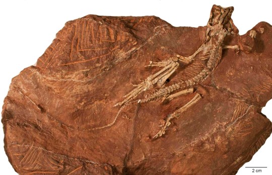
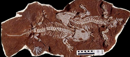
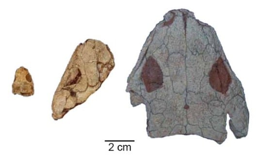
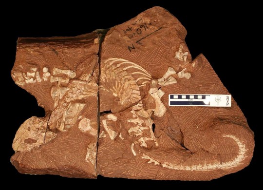
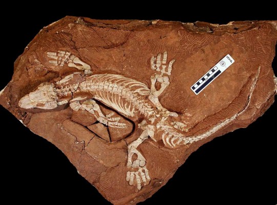
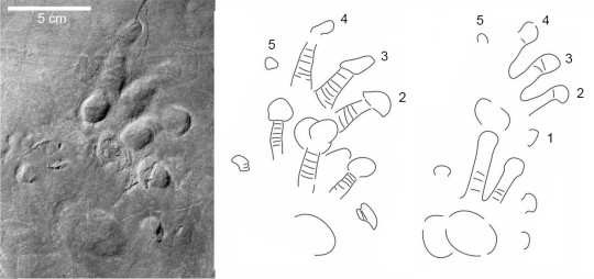
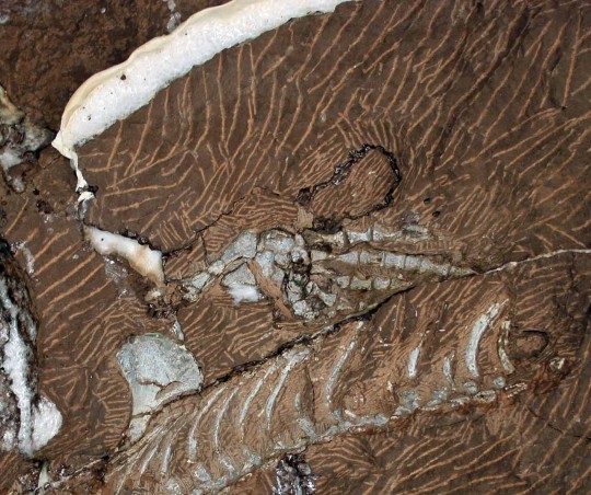
The formal publication of some of the Bromacker discoveries took more time to complete than others, and our most recently pubished fossil, Martensius bromackerensis, holds the record in that regard. Four nearly complete specimens of Martensius were collected from the Bromacker quarry between 1995–2006. The first, discovered by Thomas Martens and his father Max, came from a jumbled pocket of fossils. Unfortunately, muddy groundwater had penetrated cracks in the subsurface of this portion of the quarry and coated and eroded bone present along these cracks. Despite this damage and the lack of a skull, we could identify the specimen as a caseid synapsid (synapsids, also known as mammal-like reptiles, are a group of amniotes whose later-occurring members gave rise to mammals).

Drawing of 1995 Martensius bromackerensis specimen. Because the specimen was collected in numerous pieces of rock, with parts of some bones exposed on apposing rocks, Scientific Illustrator Kevin Dupuis (UTM) had to first draw the bones exposed on each piece and then assemble all of the drawings digitally. Dotted lines indicate bone impression in the rock. Arrows point to healing scars from two fractures in the last right rib. Additional healing scars can be seen in preceding ribs. This animal apparently survived a serious injury. Modified from Berman et al., 2020.

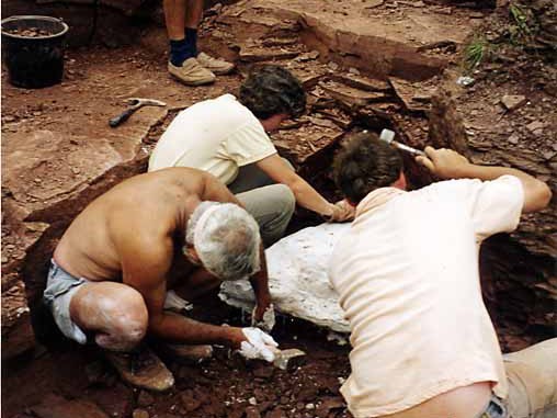
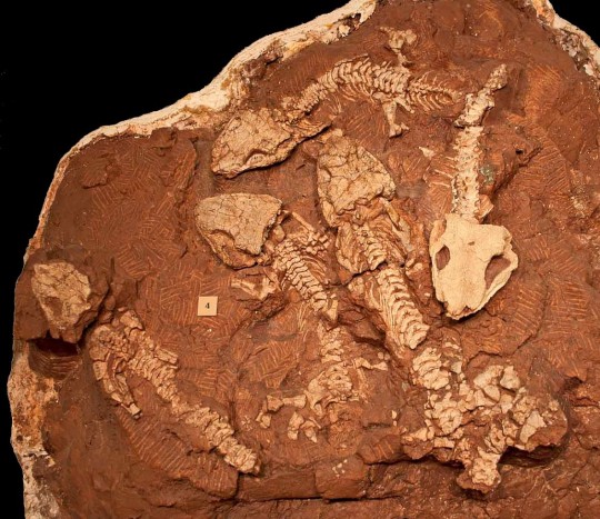
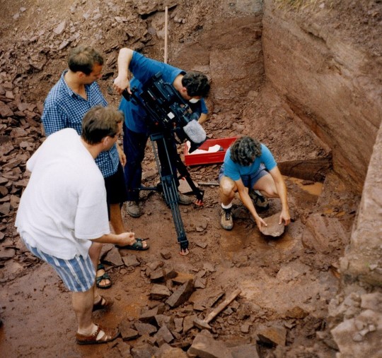
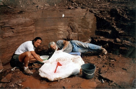
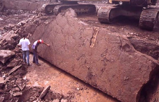
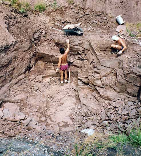
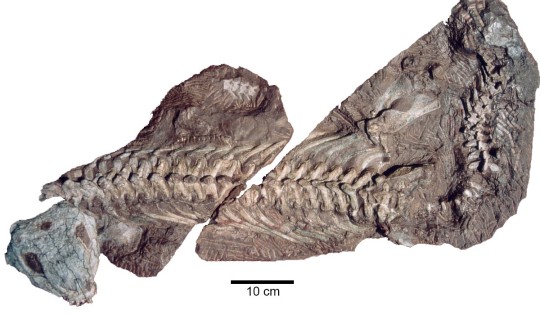
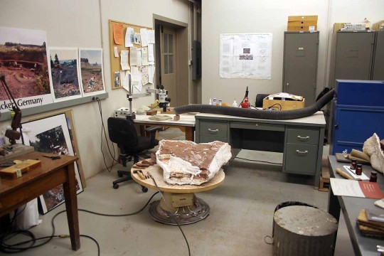
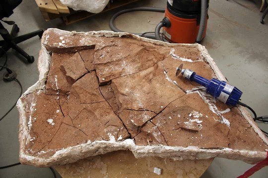
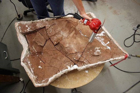
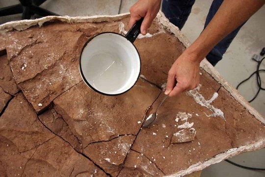
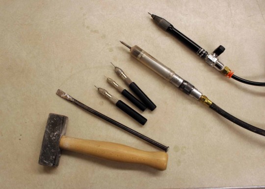
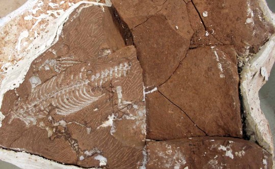
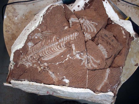
The next specimen was discovered in 1999 by Georg Sommers (Preparator, Museum der Natur, Gotha), who prepared the fossil. It consists of a vertebral column, ribs, some limb bones, and a few scattered skull elements. Unfortunately, a more complete skull was needed to allow for comparison to other caseids, some of which are based only on skull material. It wasn’t until the discovery of two more specimens in 2004 and 2006 by Stuart Sumida and Dave Berman, respectively, that the long sought-after skull was found. Preparation of these specimens took a long time due to their size and the considerable amount of rock covering the bones in some of the blocks. My promotion to Collection Manager in 2005 left me with considerably less time to prepare fossils. Other preparators were asked to help with the preparation at both Carnegie Museum of Natural History (CMNH, Dan Pickering and Tyler Schlotterbeck) and in Dr. Robert Reisz’s lab at the University of Toronto at Mississauga (Diane Scott and Nicola Wong Ken). Robert was originally slated to lead the study, but other commitments prevented him from working on it, so Dave took over.

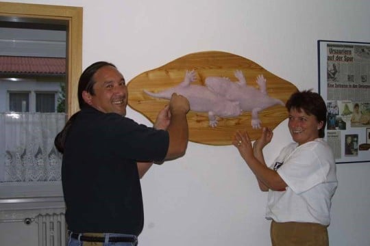
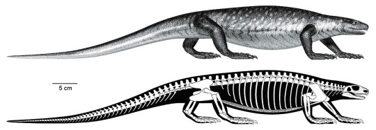
Besides preparation, the scientific study and publication of the specimens required illustrations and photographs, most of which were done by Diane, Nicola, and Kevin. Andrew McAfee (Scientific Illustrator, CMNH) made skeletal and flesh reconstructions of the animal, as well as an illustration of two Martensius in their ancient habitat (see The Bromacker Fossil Project Part III for a link to this illustration). All of this effort was worth it, however, because besides adding to the diversity of the Bromacker vertebrate fauna, Martensius has an unusual life history.

Juvenile specimen of Martensius bromackerensis. Image digitally assembled by the author from two photographs (skull and body) taken by Diane Scott in 2013. The skull, shown in ventral aspect, is incomplete and eroded on its dorsal surface.

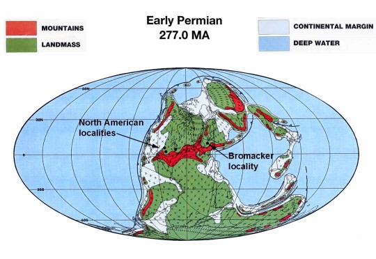
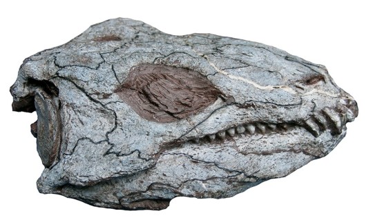
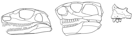
Caseid synapsids are a diverse, long-lived group known from the Late Pennsylvanian–Middle Permian epochs (~300–259 million years ago) of Europe, Russia, and the USA, and, with one exception, all are adapted to eating plants (herbivorous). The most advanced caseids (such as the enormous Cotylorhynchus romeri) have ridiculously small skulls when compared to those of carnivores, spatulate (spoon-shaped) teeth tipped with small tubercles (cuspules) for cropping vegetation, and huge, barrel-shaped ribcages to support a large gut for fermenting cellulose-rich plants. The exception is the earliest known (Late Pennsylvanian epoch, ~300 million years ago) caseid, Eocasea martini, represented by a single, incomplete juvenile specimen from Kansas. The teeth of Eocasea are small and conical, which indicate that it most likely ate insects. Because it’s skull and ribcage are of normal size, in contrast to juveniles of Martenius, Eocasea probably ate insects throughout its life.

Reconstruction of the skull of Martensius bromackerensis (left) from the Early Permian (~290 million years ago) Bromacker quarry, Germany, and the more advanced caseid Ennatosaurus tecton (skull, middle and skull fragment with cuspule-tipped teeth, right), from the Middle Permian (~263 million years ago) of Russia. Skull reconstruction of Martensius made by Diane Scott and modified from Berman et al., 2020. Ennatosaurus skull reconstruction and jaw fragment drawing modified from Maddin et al., 2008.

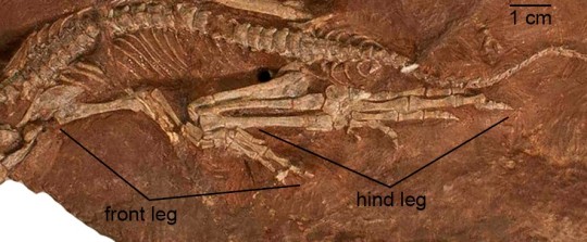
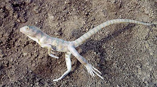
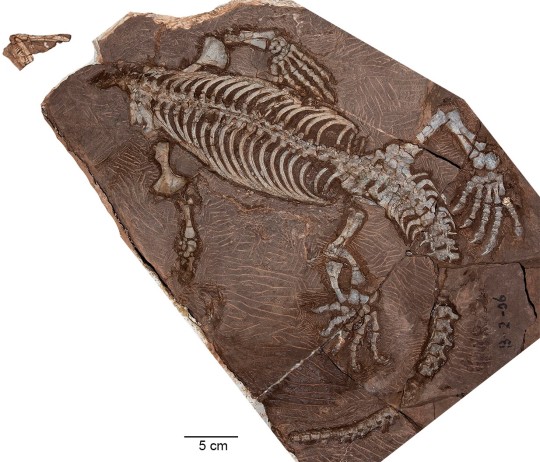
Martensius has a modestly expanded ribcage and a small skull, suggesting that it was herbivorous. Furthermore, the feet of Martensius, like those of other caseids in which the feet are known, are large, with massive, elongated, strongly recurved claws. Martensius also has a well-supported hip region that may have enabled it to rise on its hind legs to reach and tear down overhead branches to feed upon.

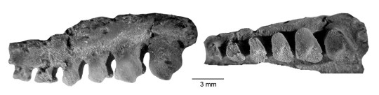
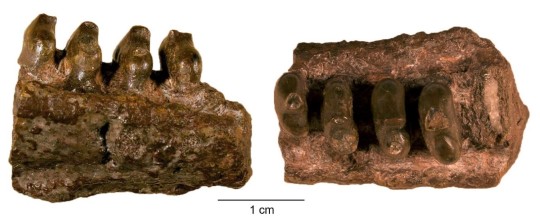
The upper and lower teeth of the adult Martensius differ from those of more advanced caseids in being triangular and lacking cuspules. The upper jaw teeth of the juvenile resemble those of the adult, but the lower jaw teeth are more numerous—31 in the juvenile compared to 25 in the adult—and surprisingly, they resemble those of Eocasea. Dave concluded that juveniles of Martensius had teeth adapted for eating insects, which were replaced by an adult dentition that would’ve been good for cropping plants and piercing insects. Remarkably, the juvenile Martensius apparently died while in the process of replacing its juvenile dentition with that of adults.

So why have different juvenile and adult dentitions? Modern animals that eat fibrous plant matter have micro-organisms called fermentative endosymbionts in their large guts, which break down difficult-to-digest plant matter via fermentation. It is assumed that early fossil plant-eaters with broad ribcages also had large guts housing fermentative endosymbionts. Prior to the discovery of Martensius, other scientists hypothesized that early herbivores acquired endosymbionts by eating herbivorous insects that already had these microbes in their guts. In Martensius, the introduction of endosymbionts apparently occurred during the juvenile, insectivorous stage of life, which set the stage for adults to add plants to their diet.

Flesh (top) and skeletal (bottom) reconstructions of Martensius bromackerensis. Illustrations by Andrew McAfee and modified from Berman et al., 2020.

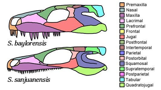
The generic name Martensius honors Thomas Martens for his discovery of vertebrate fossils at the Bromacker quarry and his perseverance in maintaining a highly successful, long-term field operation resulting in the discovery and publication of the exceptionally preserved Bromacker fossils. Bromackerensis refers to the Bromacker quarry, the only locality from which this species is known.

Stay tuned for my next post, which will feature some terrestrial dissorophoid amphibians.

For those of you who would like to learn more about Martensius, here’s a link to the 2020 Annals of Carnegie Museum publication in which it was described.

Amy Henrici is Collection Manager in the Section of Vertebrate Paleontology at Carnegie Museum of Natural History. Museum employees are encouraged to blog about their unique experiences and knowledge gained from working at the museum.

Keep Reading

The Bromacker Fossil Project Part IX: The Dissorophoid Amphibians Tambachia, Rotaryus, and Georgenthalia, Capable Travelers

Filed Under: Blog Tagged With: Amy Henrici, dinosaurs in their time, fossils, Museum from Home, paleontology, Science News, Section of Vertebrate Paleontology, Vertebrate Paleontology

July 15, 2020 by wpengine

The Bromacker Fossil Project Part VII: Eudibamus cursoris, the Original Two-legged Runner

New to this series? Read The Bromacker Fossil Project Part I, Part II, Part III, Part IV, Part V, and Part VI. 

Holotype specimen of Eudibamus cursoris, the most complete bolosaurid reptile known. Photo by the author, 2013.

Stuart Sumida discovered some small bones in the Bromacker quarry in 1993, the same year that the holotype skeleton of Diadectes absitus was found. Dave Berman told me that when Stuart showed them to him, he couldn’t see anything because they were so small. Upon closer examination, Dave, Stuart, and Thomas Martens identified them as those of the captorhinomorph reptile Thuringothyris mahlendorffae. Thomas’ wife Stefani, whose maiden name is Mahlendorff, discovered the first specimen in the Bromacker in 1982, and Thomas and a colleague named it in her honor in a 1991 publication.

The fossil was exposed in several pieces of rock, which Thomas shipped to Carnegie Museum of Natural History (CMNH) along with the large block of rock containing Diadectes. I didn’t prepare the specimen until several years later, as other projects, including the Diadectes, overshadowed it. Once I began working on it, though, Dave and I realized that it was not Thuringothyris. Indeed, we had no idea what type of animal it was, and our puzzlement grew as I exposed more of it. The identity wasn’t revealed until I had uncovered some very unusual, tiny teeth, which under the high magnification of the preparation microscope appeared to have a bulbous cusp towering over a basin. They looked vaguely familiar to me, but because I couldn’t immediately put a name on them, I rushed to get Dave from his office. Once Dave saw the teeth, he realized that the specimen was a new genus and species in the rare, enigmatic reptile group Bolosauridae.

Tiny teeth of a bolosaurid reptile, Bolosaurus striatus, in lateral (side; left) and occlusal (chewing surface; right) views. The specimen is in the CMNH Vertebrate Paleontology collection. Photos by Spencer Lucas (Research Associate, CMNH).

Until the discovery of Eudibamus cursoris, bolosaurids were represented in the fossil record by two genera, Bolosaurus and Belebey, which were based mainly on poorly preserved skull and fragmentary jaw fossils from Texas and Russia, respectively. Even though bolosaurids had been known since 1878, their relationship to other reptiles was not well understood. The nearly complete anatomy of Eudibamus allowed our team to determine that bolosaurids are the oldest member of the ancient group of reptiles called Parareptilia. This group has no living relatives, except possibly for turtles, a hypothesis that is highly debated by scientists.

Eudibamus cursoris fossil
Closeup of front and hind legs of Eudibamus. The hind leg, folded upon itself, is considerably longer than the front leg. Photo by the author, 2013.

When our study of the fossil began, we realized that Eudibamus was very different than other reptiles from that time. Proportions of the limbs and positions of the articulation surfaces on the upper and lower hind leg bones indicated that, in terms of posture, Eudibamus resembled a bow-legged human with a bad back instead of a typical sprawling reptile on four legs. It could stand and locomote on its hind legs in an upright posture (bipedal) with its legs held close together and in the same plane (parasagittal).

Dave was in constant phone communication with team member Dr. Robert Reisz (Professor, University of Toronto at Mississauga). One day Robert called Dave to ask if all the tail had been exposed, because he learned that modern lizards that are able to run bipedally have a long tail to help maintain their balance. The specimen was in Dave’s office and he immediately uncovered more of the tail and then let me finish the task. The tail was indeed very long and extended close to the edge of the block, which I had previously reduced in size. Additionally, we determined that the third, fourth, and fifth toes of the hind foot also were greatly elongated through lengthening of some of the individual toe bones, and that the first and second toes were extremely shortened by the reduction in size of individual toe bones. We hypothesized that when Eudibamus ran bipedally, it would rise on its toes, so that only the tips of the third, fourth, and fifth toes would contact the ground.

Drawing of the hind leg of Eudibamus cursoris (left) and the roughly contemporaneous reptile Captorhinus (right). Leg drawings are scaled to the same torso length of the whole animal. Illustrations of the animals are not to scale. Hind leg drawings are modified from Berman et al., 2000 and animal illustrations are from Wikimedia Commons.

Eudibamus occurred at least 60 million years before other bipedal, parasagittally-running reptiles appeared in the fossil record. This is reflected in its scientific name, which is derived from the Greek “eu,” meaning original or primitive, and “dibamos,” meaning on two legs. “Cursoris” is Latin, meaning runner. Examples of other reptiles using this locomotion mode are the dinosaurs Allosaurus fragilis and Tyrannosaurus rex, which you can view in CMNH’s Dinosaurs in Their Time exhibition.

So, what was the advantage of being able to run bipedally instead of running on all four legs? Lengthening the hind leg and foot would greatly increase stride length, especially if only the tips of the toes contacted the ground, which is an efficient way to increase speed. Eliminating arm to ground contact while running removes forelimbs from the path of the long-striding hind legs. The bulbous teeth and jaw structure of Eudibamusindicate that it was herbivorous. It seems likely, then, that Eudibamus used its ability to sprint to avoid becoming a tasty meal for a pursuing predator.

Eudibamus cursoris illustration
Peter Mildner (exhibit preparator at the Museum der Natur, Gotha) made a surprise visit to the Bromacker one afternoon to show us a model of Eudibamus cursoris he’d made. This image shows the model in the present day Bromacker quarry, part of the region it inhabited 290 million years ago. Photo by the author, 2006.

One of our laments is that a fossil trackway preserving Eudibamus walking quadrupedally and then switching to a bipedal gait has yet to be found.

Next time you are at CMNH, make sure you see the cast of the fossil skeleton and a model of Eudibamus that are exhibited in the Fossil Frontiers display case in CMNH’s Dinosaurs in Their Time exhibition. Stay tuned for my next post, which will feature the herbivorous mammal-like reptile Martensius bromackerensis.

For those of you who would like to learn more about Eudibamus, here is a link to the 2000 Science publication in which it was described: https://science.sciencemag.org/content/290/5493/969.

Amy Henrici is Collection Manager in the Section of Vertebrate Paleontology at Carnegie Museum of Natural History. Museum employees are encouraged to blog about their unique experiences and knowledge gained from working at the museum.

Keep Reading

The Bromacker Fossil Project Part VIII: Martensius bromackerensis, Honoring a Colleague

Filed Under: Blog Tagged With: Amy Henrici, dinosaurs in their time, Museum from Home, Science News, The Bromacker Fossil Project, Vertebrate Paleontology

June 24, 2020 by wpengine

The Bromacker Project Part VI: Seymouria sanjuanensis, the Tambach Lovers

New to this series? Read The Bromacker Fossil Project Part I, Part II, Part III, Part IV, and Part V.

Seymouria sanjuanensis fossils
Two exquisitely preserved, nearly complete adult skeletons of Seymouria sanjuanensis that were discovered in the Bromacker quarry in 1997. Photo by Dave Berman.

At lunchtime on the last day of the 1997 field season, Thomas Martens discovered the two exquiste specimens shown above, the only fossils found that year. Thomas had uncovered a piece of the hip region with some attached vertebrae that resembled, once again, those of the ancient amphibian Seymouria. Because our work time was limited, we estimated the length of the specimen and rushed to extract it from the quarry. When we flipped the block over, a few pieces of rock fell out, revealing a series of vertebrae of a second individual in the block. We were thrilled to learn that Thomas had discovered two specimens of Seymouria. We put the rock pieces back in place and quickly finished plastering the block. There was just enough time for Dave, Stuart Sumida, and I to return to our hotel, clean up, quickly pack, and meet Thomas, his family, and his fossil preparator Georg Sommer for a celebratory dinner. What a great way to end the field season.

Working in tight quarters to quickly extract the Seymouria specimens discovered at lunchtime on the last day of the field season. Clockwise from right: Georg Sommer, Dave Berman, and the author. Photo by Stuart Sumida, 1997.

Seymouria had already been known from the Bromacker quarry. Thomas had discovered and identified two skulls in 1985, fossils he brought with him when he came to Carnegie Museum of Natural History (CMNH) in 1993 to study for six months with Dave Berman under a CMNH-financed fellowship. Both skulls were of juvenile individuals. Of the two known species of Seymouria, Dave and Thomas were excited to discover that the Bromacker skulls were nearly identical to those of Seymouria sanjuanensis. The 1997 lunchtime discovery of the two complete adult specimens confirmed the identification of the Bromacker Seymouria as S. sanjuanensis.

The first discovered species of Seymouria was Seymouria baylorensis, from near Seymour, Baylor County, Texas, from which its name was derived. Seymouria sanjuanensis was first found in San Juan County, Utah, by Dave Berman and the field team he was leading as a graduate student at the University of California, Los Angeles. Dave’s advisor, Dr. Peter Vaughn, named it Seymouria sanjuanensis in reference to the county of discovery. Another discovery of five specimens of this species preserved together was made by Dave in New Mexico in 1982.

Comparison of the skulls of Seymouria baylorensis (top) and S. sanjuanensis (bottom). The individual bones of the skull are color coded. Skulls scaled to same size. Image from Wikimedia Commons.

Seymouria baylorensis is geologically younger than S. sanjuanensis and has a more robust skull, larger and fewer teeth of variable size, and a subrectangular postorbital bone compared to the chevron-shaped postorbital of S. sanjuanensis.

Seymouria is considered a terrestrial amphibian that only returned to water to breed. Its strongly built skeleton provided the support needed to move on land. With its numerous, slender, pointed teeth, S. sanjuanensis most likely ate insects and small land-living vertebrates. We know that the Bromacker Seymouria didn’t consume fish, because not a single fish fossil, scrap of fish fossil, or fish coprolite (fossil poop) has ever been found at the Bromacker quarry. Study of the rock deposits preserving the fossils at the Bromacker indicate a lack of permanent water, which would explain the absence of fish.

Growth series of skulls of Seymouria sanjuanensis from the Bromacker Quarry showing (left to right) early juvenile, late juvenile, and adult growth stages. Photos by the author, 2006.

Conditions for breeding must have been favorable in the Tambach Basin, the ancient basin where sediments preserving the Bromacker fossils accumulated, because several juvenile specimens of Seymouria are known. The smallest is a skull measuring about ¾ of an inch long. In a study led by our colleague Josef Klembara (Comenius University, Slovak Republic), we determined that the smallest individual was post-metamorphic—in other words, no longer a tadpole—based on the presence of certain ossified bones in the skull. In tadpoles, these skull elements are cartilaginous; that is, they haven’t yet turned to bone.

Seymouria sanjuanensis fossils
Five skeletons of Seymouria sanjuanensis preserved together were discovered in north central New Mexico by Dave Berman in 1982. These specimens are on display in CMNH’s Benedum Hall of Geology, in the “What is a Fossil?” case. Photo by the author, 2013.

The discovery in Germany of the same species of Seymouria previously known only from New Mexico and Utah has important implications in terms of paleobiogeography (the study of the distribution of species in space and time). At the time S. sanjuanensis was alive, the continents were merged to form the supercontinent Pangaea. The presence of S. sanjuanensis across Pangaea, north of a roughly east-west trending mountain range, indicates that climatic or physical barriers (e.g., deserts, inland seas, mountain ranges) didn’t prevent its dispersal.

Map showing the arrangements of the continents in the Early Permian. The locality where Seymouria occurs in present-day New Mexico, Texas, and Utah and the Bromacker locality in present-day Germany are indicated. Map modified from Scotese, 1987.

The two Seymouria specimens preserved together were a big hit in the local region in Germany. Museum der Natur (MNG) exhibit preparator Peter Mildner nicknamed them the “Tambacher Liebespaar” (“Tambach Lovers”) after a painting entitled “Gothaer Liebespaar” (“Gotha Lovers”) on exhibit in the Herzogliches Museum of the Stiftung Schloss Friedenstein (also the parent organization of MNG). This name caught on and is fondly used by our German friends and colleagues. Peter even made a fleshed-out model of the two Seymouria specimens in their death pose. The proprietor of the hotel in which we stayed hung a copy of the model of the Tambach Lovers and a framed collage of newspaper articles featuring the Bromacker on a wall in one of the hotel rooms, which she named the “Präparation Suite” (i.e. “Preparation Suite” in reference to the preparation of fossils). I often stayed in this room.

The painting entitled “Gothaer Liebespaar” (“Gotha Lovers”), which is on display at Herzogliches Museum of the Stiftung Schloss Friedenstein, Gotha, Germany. Image from Wikimedia Commons and provided by Thomas Martens.

Tambach Lovers postcard
Postcard showing the Tambach Lovers. The postcard was made for and sold by the Museum der Natur, Gotha. Photo of the postcard by the author, 2020.

Stuart Sumida (left) and Heike Scheffel, proprietor of the Hotel Wanderslaben where we stayed (right), with the model of the Tambach Lovers in the “Präparation Suite.” The framed collage to the right of the model holds newspaper articles featuring the Bromacker project. Photo by the author, 2003.

A cast of the Tambach Lovers specimen and a model of Seymouria sanjuanensis are exhibited in the Fossil Frontiers display case in CMNH’s Dinosaurs in Their Time exhibition. Be sure to look for them once the museum re-opens. And stay tuned for my next post, which will feature the unusual bipedal reptile Eudibamus cursoris.

For those of you who would like to learn more about Seymouria sanjuanensis, here is a link to the publication describing the 1997 specimens: https://www.tandfonline.com/doi/abs/10.1671/0272-4634(2000)020%5B0253%3AROSSSF%5D2.0.CO%3B2.

Amy Henrici is Collection Manager in the Section of Vertebrate Paleontology at Carnegie Museum of Natural History. Museum employees are encouraged to blog about their unique experiences and knowledge gained from working at the museum.

Keep Reading

The Bromacker Fossil Project Part VII: Eudibamus cursoris, the Original Two-legged Runner

Filed Under: Blog Tagged With: Amy Henrici, fossils, Museum from Home, Science News, The Bromacker Fossil Project, Vertebrate Paleontology

June 10, 2020 by wpengine

The Bromacker Project Part V: Orobates pabsti, Pabst’s Mountain Walker

New to this series? Read The Bromacker Fossil Project Part I, Part II, Part III, and Part IV. 

In 1995, my first year of field work at the Bromacker quarry, Stuart Sumida discovered a fossil that we initially thought was that of the amphibian Seymouria, based on the size and shape of the exposed vertebrae. This tentative identification made sense, because before our collaboration began, Thomas Martens had discovered in the Bromacker a skull of Seymouria, a creature known from localities in the USA. Months later, while I was preparing the specimen, Dave Berman and I realized the fossil wasn’t Seymouria, and that it belonged to the same unnamed animal that Thomas had collected a partial skeleton of before our collaboration began.

image of orobates pabsti
Specimen of Orobates pabsti collected in the 1995 field season. We determined that it is a juvenile. Photo by Dave Berman.

In the 1998 field season I discovered a third specimen, which is by far the most spectacular fossil that I have ever discovered. I found it towards the end of the field season when I pried up a piece of rock from the quarry floor. Upon turning over the rock piece, I saw an articulated foot preserved in it. I couldn’t believe my eyes! I knew that at the Bromacker if an articulated foot was found, the rest of the articulated skeleton should be attached to it. The problem was, we didn’t know if I had discovered a front or a hind foot, so we weren’t sure how the specimen was oriented in the quarry and whether it penetrated the nearby rock wall. Dave carefully lifted another piece of rock and thought the bones exposed in it were part of the shoulder girdle. Unfortunately, closer examination revealed that it was a piece of skull roof—another lobotomy—but, lacking x-ray vision, this is how we find fossil bone at the Bromacker. The good news was that the fossil specimen appeared to parallel the quarry wall.

image
A film crew from the regional MDR television station visited us early on the day of my discovery to interview Dave and Thomas. The discovery was made after they left, so Thomas immediately notified them. They returned and recorded a reenactment of my discovery. The piece of rock I am holding contains the foot. The rest of the fossil lies in the low mound of rocks in front of me. Photo by Dave Berman, 1998.

image
Dave and Stuart finish plastering the block. The red flag is a north arrow to indicate the orientation of the block in the quarry. Photo by the author, 1998.

Dave, Stuart, Thomas, then-graduate student Richard Kissel (University of Toronto, Mississauga), and I named the animal Orobates pabsti, which is from the Greek “oros,” meaning mountain, and “bates,” meaning walker, in reference to the Bromacker fossil environment being an intermontane basin. “Pabsti” is in honor of Professor Wilhelm Pabst for his pioneering work on the Bromacker fossil trackways.

We determined that Orobates is very closely related to Diadectes, and like Diadectes, was herbivorous. Orobates differs from Diadectes and other diadectomorphs in the group Diadectidae in a number of features, some of which are as follows: spade-shaped cheek teeth that are oriented on the jaw at an angle of 30–40° to the jaw line, rather than being close to 90°; narrower and shorter vertebral spines; 26 vertebrae between the head and hip (Diadectes has 21); proportions and shapes of individual toe bones; and digit (finger or toe) length.

image of orobates pabsti
Holotype specimen of Orobates pabsti, the specimen collected in 1998. If a series of specimens exists of a new species, then the specimen that best represents the species is designated as the holotype. If only one specimen is known, it becomes the holotype by default. Photo by Dave Berman.

The Bromacker has long been famous for its exquisitely preserved fossil trackways. Identification of the particular fossil animal that made a given trackway is almost always very difficult, because body fossils often lack completely preserved hands and feet and typically are not found in association with trackways. As a result, trackways are given their own set of names, called ichnotaxa (“ichno” means track or footprint), which are typically referred to major groups of animals instead of individual species. The Bromacker is unique, however, because nearly completely preserved body fossils occur in a rock unit above the trackways, indicating they are very nearly contemporaneous. Five ichnotaxa are known from the Bromacker, and one of them, Ichniotherium, has been attributed to Diadectidae.

image
A large slab of rock being inspected for trackways shortly after it was unearthed in the commercial rock quarry. The polygonal patterns in the rock are mudcracks. Photo by the author.

Graduate student and trackway expert Sebastian Voigt (now Director at Urweltmuseum GEOSKOP, Burg Lichtenberg, Germany) often visited us at the Bromacker. In 2000, a time when Diadectes was the only known Bromacker diadectid, Sebastian and his advisor Hartmut Haubold (now emeritus at Martin Luther University Halle-Wittenberg, Germany) proposed that Ichniotherium cottae made two track types, designated as A and B, that differed according to the speed at which the trackmakers moved. This contrasted previous studies that proposed three species of Ichniotherium at the Bromacker.

Once the skeletal anatomy of Orobates became known, Sebastian realized that there were two species of Ichniotherium, and they were made by Diadectes and Orobates, respectively. He invited Dave and me to co-author a paper to present this hypothesis. We supplied Sebastian with information about skeletal differences between Diadectes and Orobates, and Sebastian used these data to firmly establish that Diadectes made Ichniotherium cottae (type B) tracks and Orobates was the trackmaker of trackways formerly identified as I.sphaerodactylum (aka I. cottae type A). Even though the makers of the trackways are now known, the ichnotaxon names are still used when referring to the trackways.

image
Photographs of trackways of Ichniotherium sphaerodactylum made by Orobates pabsti (top) and Ichniotherium cottae made by Diadectes absitus (bottom). Modified from Voigt (2007).

In Diadectes, the fifth digit of the hind foot is relatively shorter than it is in Orobates, which can be seen in the tracks of I. cottae and I. sphaerodactylum, respectively. Furthermore, in I. cottae trackways, the hind foot track overlaps the track of the front foot, whereas in I. sphaerodactylum the hind foot track typically doesn’t overlap the front foot track. This is because Diadectes has less vertebrae between the head and hip (21 vertebrae) than Orobates (26 vertebrae) does.

image
Front and hind foot track pair of Ichniotherium sphaerodactylum. Track made by the front foot is above the hind foot track. Digits 1–5 indicated. Modified from Voigt, 2007.

image
Front and hind foot track pair of Ichniotherium cottae. Track made by the front foot is above the hind foot track. Digits 1–5 indicated. Notice that the hind foot track overlaps the front foot track. Drawings are of different specimens than the one photographed. Modified from Voigt, 2007.

A cast of the holotype skeleton of Orobates pabsti is exhibited in the Fossil Frontiers display case in Carnegie Museum of Natural History’s Dinosaurs in Their Time exhibition. Be sure to look for it once the museum re-opens. And stay tuned for my next post, which will feature the amphibian Seymouria sanjuanensis.

For those of you who would like to learn more about Orobates, you can access the abstract here or contact Amy Henrici here. The publication on the track-trackmaker association can be found here.

Amy Henrici is Collection Manager in the Section of Vertebrate Paleontology at Carnegie Museum of Natural History. Museum staff, volunteers, and interns are encouraged to blog about their unique experiences and knowledge gained from working at the museum.

Keep Reading

The Bromacker Fossil Project Part VI: Seymouria sanjuanensis, the Tambach Lovers

Filed Under: Blog Tagged With: Amy Henrici, Museum from Home, Science News, Section of Vertebrate Paleontology, The Bromacker Fossil Project, Vertebrate Paleontology

June 4, 2020 by wpengine

The Bromacker Project Part IV: Diadectes absitus, A Project-Saving Fossil

New to this series? Read The Bromacker Fossil Project Part I, Part II, and Part III.

This post will be the first of a series focusing on notable fossil animals discovered in the Bromacker quarry. I selected Diadectes absitus, a member of the ancient group Diadectomorpha, to present first because, had it not been discovered in the first year of the collaborative field work, the project might not have continued.

Dave Berman and his colleague Stuart Sumida (California State University, San Bernardino) joined Thomas Martens for five weeks of field work in the summer of 1993. They dug a quarry over six feet deep in their search for fossils, while working in a mix of hot and humid or near freezing temperatures, with plenty of rain. It wasn’t until the second-to-last day of the field season that the Diadectes specimen was discovered. By then, as Dave later told me, he was so discouraged by the lack of fossils that he assumed this would be his first and last field season in Germany. I should mention that Stuart had previously uncovered a few small vertebrae, but because the vertebrae resembled an animal described from the Bromacker in 1991, the team was not very excited about the discovery. They couldn’t have been more wrong in their field identification of the vertebrae, however, but more on that in a later post.

The 1993 quarry shortly before discovery of Diadectes absitus. Pictured are Stuart Sumida standing in the quarry and Thomas Martens crouched to his right. The Diadectes fossil was found in the corner opposite Stuart’s left shoulder, which is out of sight in this image. Photo by Dave Berman, 1993.

The team’s collective attitude changed when Stuart knocked off a chunk of bone-bearing rock from a bench in the quarry corner while shoveling away rock rubble. Careful examination of the fragment revealed part of the top of a roughly five-inch-long skull. We have since joked that Stuart gave it a lobotomy. While collecting the large block of rock containing the remainder of the skull, another piece of rock popped off the the edge of the block adjacent to the quarry wall. This piece had vertebrae in it. The team then realized that only the front portion of the animal was in the block freed from the quarry. The rest of the fossil skeleton remained in the quarry wall. Thomas later excavated the rest of the specimen and shipped it separately to Carnegie Museum of Natural History (CMNH).

You can watch Dave and Stuart excavate the fossil-bearing block by clicking on the video link at the end of this post.

Based on the shape of both the exposed teeth in the broken skull and the exposed vertebrae, Dave and Stuart were able to identify the fossil animal as the genus Diadectes. Thomas had already collected a juvenile skull and other bones of Diadectes before his collaboration with Dave, but the specimen discovered in 1993 was by far the most complete and best preserved.

Skeleton of Diadectes absitus. Some of the limb bones are preserved on the underside of the block. Photo provided by Thomas Martens.

Once my preparation of the specimen was completed, which took a little over a year, Dave, Stuart, and Thomas begin their detailed study and description of the fossil. They determined that it represented a new species, which they named Diadectes absitus. “Absitus” is Latin for distant or far, in reference to the species being the first occurrence of Diadectes outside of North America. The generic name Diadectes was coined in 1878 by the famous paleontologist Edward Drinker Cope, and is a combination of the Greek “dia,” meaning crosswise, and “dēktēs,” meaning biter, in reference to its broad teeth. Other species of Diadectes occur in similar-aged rocks in the American southwest, and a few specimens are known from the Tri-State area of Pennsylvania, Ohio, and West Virginia.

Diadectes is a member of the group Diadectomorpha, which has oscillated between being considered a member of Amphibia or Amniota. Amphibians lay their eggs in water, which then hatch into tadpoles that later undergo metamorphosis. Today this group includes frogs, salamanders, and caecilians (limbless, worm-like burrowing amphibians). In contrast, amniotes either lay their eggs on land, like reptiles and birds do today, or the embryo develops sufficiently in the mother for live birth, as in most mammals. Except in rare cases, the type of developmental pathways of fossil animals cannot be determined because they are rarely preserved with their eggs or fetuses. Paleontologists instead study a variety of preserved features to determine group membership. As an example, amphibians typically have four fingers, whereas amniotes generally possess five.

Diadectes and its close relatives were herbivorous, that is, they ate plants. Their spatulate, incisor-like front teeth project forwards and were adapted for cropping vegetation. Longitudinal, parallel striations on their broad cheek teeth suggest that Diadectes could move its lower jaw fore and aft to grind plant matter against its upper jaw teeth, a motion called propalinal.

Skull of Diadectes absitus in right lateral (= right side) aspect. Notice the forward-angled front teeth and the bulbous cheek teeth. A black pen was used to mark the boundaries of individual bones in the skull, which aided study of the animal. Modified from photo provided by Thomas Martens.

The presence of an enlarged torso and teeth adapted for grinding tough vegetation are evidence that Diadectes absitus likely consumed a diet of high-fiber plants. Animals that eat high fiber plants, such as cows, have enlarged torsos framed by a rounded rib cage to hold large guts for processing plant cellulose through fermentation by microorganisms.

Diadectes absitus lived at a time when herbivores were just beginning to evolve. One of the oldest known herbivores is the diadectomorph Desmatodon hollandi, which lived about 305 million years ago, whereas Diadectes absitus lived roughly 290 million years ago. We discovered a surprisingly high number of herbivores at the Bromacker.

Teeth of one of the oldest known herbivores, the diadectomorph Desmatodon hollandi. This specimen was discovered in Pitcairn, PA by Percy E. Raymund (Assistant Curator, Section of Invertebrate Paleontology) in 1907 and named in honor of Dr. William Holland, the second Director of CMNH. The teeth of Desmatodon are very similar to those of Diadectes absitus. Photo by the author, 2018.

A cast of the skeleton of Diadectes absitus is exhibited in the Fossil Frontiers display case in the Dinosaurs in Their Time exhibition. Be sure to look for these once the museum re-opens. And stay tuned for my next post, which features another diadectomorph, Orobates pabsti.

Photograph of a model of Diadectes absitus made by the Museum der Natur, Gotha exhibit preparator Peter Mildner. Photo provided by Thomas Martens.

Dave and Stuart excavate the fossil-bearing block (video)

Amy Henrici is Collection Manager in the Section of Vertebrate Paleontology at Carnegie Museum of Natural History. Museum employees are encouraged to blog about their unique experiences and knowledge gained from working at the museum.

Keep Reading

The Bromacker Fossil Project Part V: Orobates pabsti, Pabst’s Mountain Walker

Filed Under: Blog Tagged With: Amy Henrici, Museum from Home, Science News, Section of Vertebrate Paleontology, The Bromacker Fossil Project

May 21, 2020 by wpengine

The Bromacker Fossil Project Part III: Fossil Preparation

New to this series? Read The Bromacker Fossil Project Part I and Part II.

Most of the important fossil discoveries from the Bromacker quarry, located in the Thuringian Forest, central Germany, were shipped to the Carnegie Museum of Natural History (CMNH) for scientific preparation. Between 1993 and 2005 I was the principal preparator of Bromacker fossils.

At CMNH the arrival of a field season’s worth of fossil crates was highly anticipated by Curator Dr. Dave Berman and myself. I’d be often notified of the crate’s early morning delivery by either a grinning security guard or shipping and receiving personnel upon my arrival at the museum. Later that day it would take a team of able-bodied staff from various departments to move the crate from the loading dock to the basement preparation lab, and to lift the plaster and burlap encased block from the crate onto a table.

photo of fossil preparation lab
My work area in the basement preparation lab. The table in the center is my main work table and is a made from a dentist chair. The blue cabinet with a hose extending from it is a dust collector, and the microscope (seen at the end of the hose) is mounted on an articulated arm to make it easier to maneuver over a block. Photo by the author, 2007.

The first step of the preparation process involved opening the block; that is, removing the top of the jacket. I’d use a cast cutter, the same tool doctors use to remove a cast protecting a person’s broken bone, to cut through the top perimeter of the plaster jacket. If all went well, the top would easily lift off the block. But if the top of the jacket stuck to the block or wedged in an undercut, I’d have to cut it into smaller pieces to remove it.

photo of fossil preparation: a block of rock with a cast cutter on top
The block collected in the 2006 field season with its top removed. Exposed bone can be seen left of center. The blue tool resting on the surface of the block is a cast cutter.

Blocks from the Bromacker quarry typically have numerous cracks coursing through them, which must be stabilized before preparation begins. The product Carbowax works well for filling cracks, because, unlike plaster, it doesn’t shrink when it solidifies. Carbowax comes as a powder, which I’d melt it in a double boiler. Before pouring the hot wax into a crack, I’d heat the surrounding rock with a heat gun so that the wax could penetrate additional cracks not visible from the surface. I’d typically repeat this process numerous times during the preparation process.

image of using a heat gun on a slab of rock in the process of fossil preparation
Using a heat gun to heat the rock before pouring wax into a crack. I’d have to carefully watch the direction I aimed the heat gun so that strands of burlap sticking out of the plaster jacket wouldn’t catch fire. Photo by Norman Wuerthele, 2007.
image
Spooning hot Carbowax into a crack. The spoon was heated beforehand so that the wax wouldn’t solidify on it. It was a delicate balance between getting the spoon hot enough so the wax stayed melted but not so hot that the spoon handle burned my hand. Photo by Norman Wuerthele, 2007.

Once the block was stabilized, I began removing rock to expose the fossil. Where thick rock covered the fossil – and it sometimes was more than six inches – I’d use a small hammer and chisel to chip away chunks of rock. As I’d get closer to the fossil, I’d switch to an airscribe, which can be likened to a miniature pneumatic jack hammer. Although fossilized bone from the Bromacker was softer than the surrounding rock, the airscribe would flake the rock from the fossilized bone, leaving behind a thin veneer of rock that I’d remove using a pin vise. I’d also use the pin vise to scrape rock from bone in tight and/or delicate areas, such as teeth. All this work was performed while looking through a microscope.

photo of seven tools used for fossil preparation
Pictured are the tools that I’d use the most when preparing Bromacker fossils. From bottom left to upper right: small hammer and chisel, three pin vises that hold a rod of tungsten carbide of varying thickness and ground to different shaped tips, and two airscribes. Photo by the author, 2007.

In the block pictured in this post, I could see some tips of some vertebral spines (these are the bumps that you feel down the midline of your back) poking from the rock surface, so I began exposing them first. Because I was working on an articulated specimen (one bone connected to the next bone), I exposed it from front to the rear by simply following one bone to the next bone.

image
The skeleton emerging from the rock—vertebrae, ribs, and the right upper arm bone (humerus) are visible. Notice also the tips of vertebral spines leading away from the exposed portion of the skeleton. The lines in the rock were made by the airscribe. The white substance along cracks is Carbowax. Photo by the author, 2007.
image
Closeup view of the right foreleg and ribs. Horizontal cracks underneath the fossil made preparation difficult, because they formed gaps underlying the bone. I had to build a dam (upper left) to contain the hot wax so that the wax would penetrate the horizontal crack underlying the bone, instead of running all over the block. Photo by the author, 2007.
image
More of the fossil skeleton is exposed, including the torso, the right foreleg, part of the left foreleg, and most of the left hind leg. Photo by the author, 2007.

Parts of the hind legs and tail were collected separate from the block, because rock pieces containing them inadvertently had been tossed on the dump pile. This occurred before the specimen had been discovered, and the bone in these pieces was covered by mud and dirt. Instead of gluing them back in the block before preparation, I prepared them individually at a table under the microscope, as it made for easier viewing. Once all the fossil had been exposed and prepared, I removed excess rock to make the block smaller and lighter weight.

image
My work on the block has been completed, except for adding some plaster bandages to the end where I had removed excess rock to make the block smaller and lighter. Photo by the author, 2007.

Dave Berman and I later transported the block to a colleague’s lab at the University of Toronto, Mississauga, Canada, where the lab staff and students completed detailed preparation and scientific illustrations of the specimen. This specimen along with several others were recently described as a new genus and species, Martensius bromackerensis, in a paper published in the Annals of Carnegie Museum. This ancient creature will be the topic of a future post. To whet your appetite, here is a link to the news release announcing the publication.

Amy Henrici is Collection Manager in the Section of Vertebrate Paleontology at Carnegie Museum of Natural History. Museum employees are encouraged to blog about their unique experiences and knowledge gained from working at the museum.

Keep Reading

The Bromacker Fossil Project Part IV: Diadectes absitus, A Project-Saving Fossil

Filed Under: Blog Tagged With: Amy Henrici, Museum from Home, Science News, Section of Vertebrate Paleontology

  • « Go to Previous Page
  • Page 1
  • Page 2
  • Page 3
  • Page 4
  • Page 5
  • Go to Next Page »

sidebar

About

  • Mission & Commitments
  • Directors Team
  • Museum History

Get Involved

  • Volunteer
  • Membership
  • Carnegie Discoverers
  • Donate
  • Employment
  • Events

Bring a Group

  • Groups of 10 or More
  • Birthday Parties at the Museum
  • Field Trips

Powdermill

  • Powdermill Nature Reserve
  • Powdermill Field Trips
  • Powdermill Staff
  • Research at Powdermill

More Information

  • Image Permission Requests
  • Science Stories
  • Accessibility
  • Shopping Cart
  • Contact
  • Visitor Policies
One of the Four Carnegie Museums | © Carnegie Institute | Privacy Policy | Terms of Use | Accessibility
Rad works here logo