• Skip to primary navigation
  • Skip to main content
  • Skip to primary sidebar

Carnegie Museum of Natural History

One of the Four Carnegie Museums of Pittsburgh

  • Visit
    • Buy Tickets
    • Visitor Information
    • Exhibitions
    • Events
    • Dining at the Museum
    • Celebrate at the Museum
    • Powdermill Nature Reserve
    • Event Venue Rental
  • Learn
    • Field Trips
    • Educator Information
    • Programs at the Museum
    • Bring the Museum to You
    • Guided Programs FAQ
    • Programs Online
    • Climate and Rural Systems Partnership
  • Research
    • Scientific Sections
    • Science Stories
    • Science Videos
    • Senior Science & Research Staff
    • Museum Library
    • Science Seminars
    • Scientific Publications
    • Specimen and Artifact Identification
  • About
    • Mission & Commitments
    • Directors Team
    • Museum History
  • Tickets
  • Give
  • Shop

Blogs from our Scientific Researchers

Carnegie Museum of Natural History is home to active research and vast scientific collections. Our scientific researchers regularly contribute to the blog at the museum.

August 18, 2020 by wpengine

How to Prepare Insect Specimens

specimens and specimen preparation tools

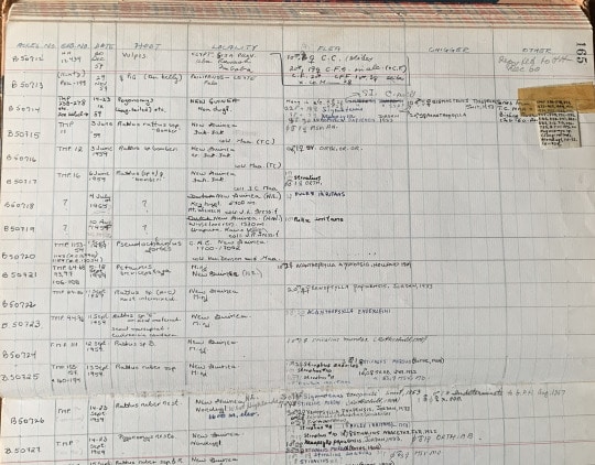
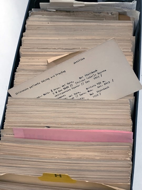
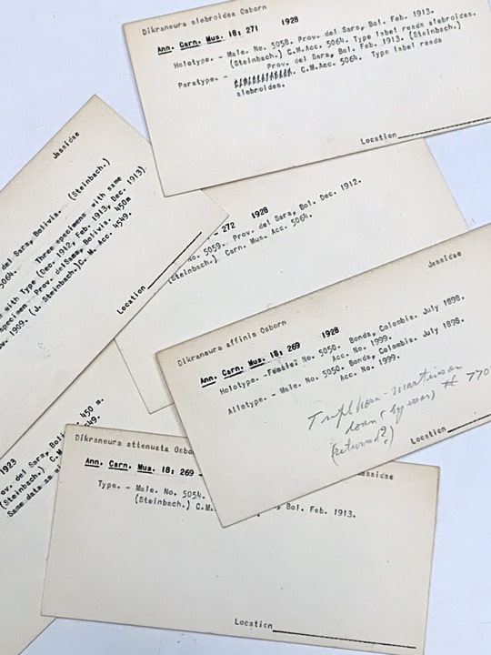
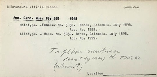
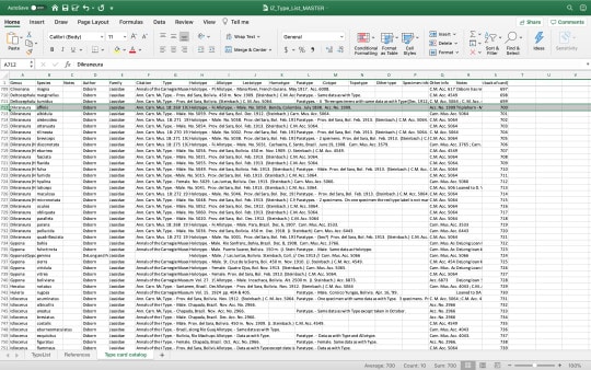
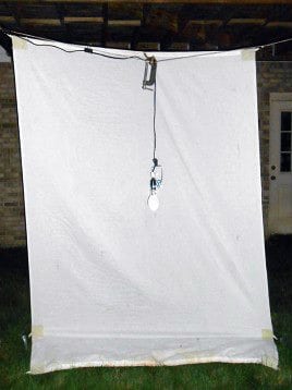
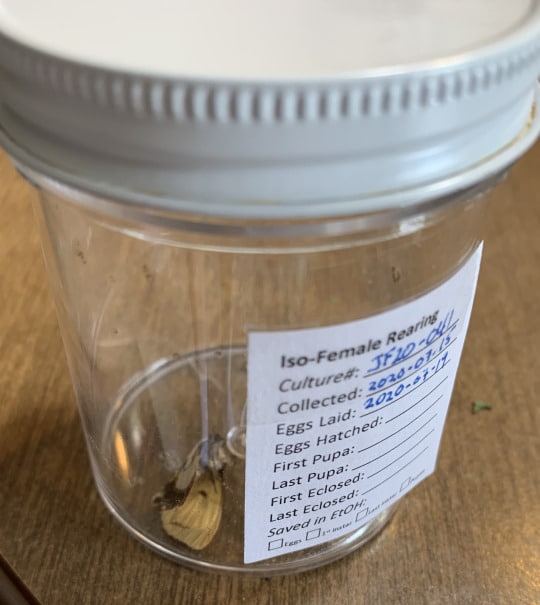
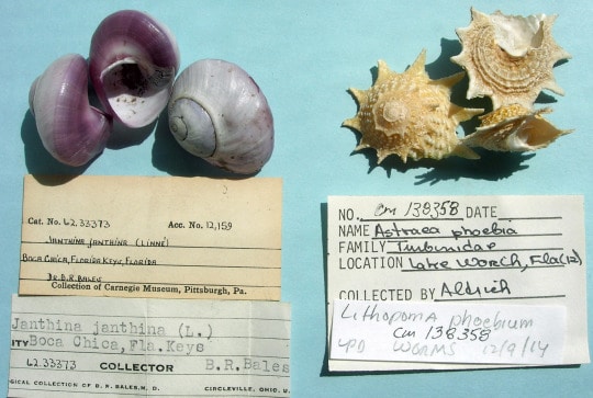
Museum collections play a significant role in helping scientists answer questions about biodiversity and in providing data that may be used for conservation studies.  Every specimen in the Invertebrate Zoology collection tells a story and all together they contribute to the story of life on Earth.  Picture it, millions of specimens prepared and labeled.  Each has a story to tell about where, when, and how it was collected.  This critically important data is also gathered when samples are collected in the field.

The next step is processing the sample and picking specimens to be prepared.  So, how do we prepare specimens?  Lepidoptera (moths and butterflies) are usually pinned in the field or preserved in the freezer and then pinned and spread in the lab.  Most non-Lepidoptera are preserved in alcohol and prepared in the lab.  Preparation techniques differ, therefore, with what is being collected.

specimens before preparation

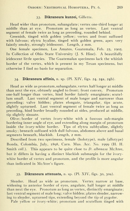
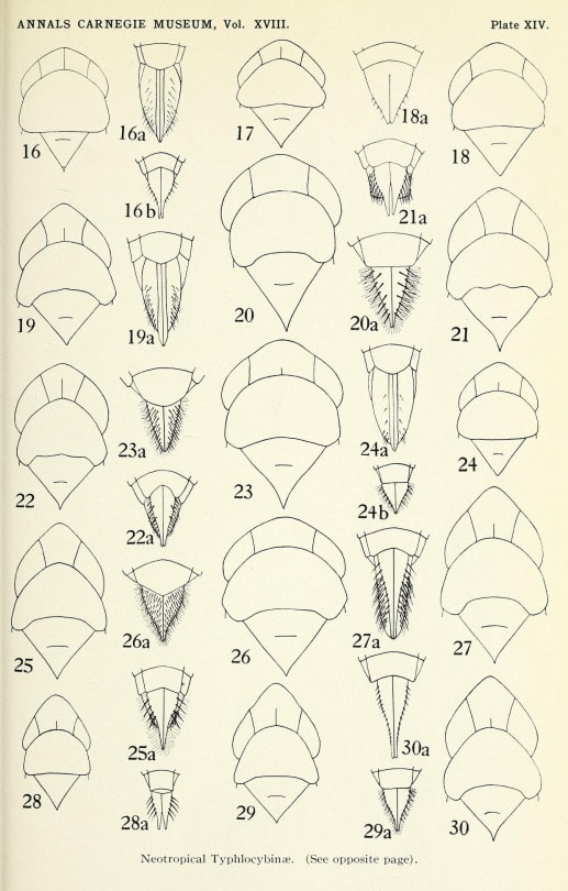
High quality scientific preparation is important, not just for aesthetic reasons but also for a specimen’s future in the service of research.  In some situations, characters on the bodies of the specimens need to be viewed under a microscope, sometimes segments need to be counted to identify a species, and more excitingly, a new species might need to be described from a series of specimens.

Handling of specimens that will be prepared needs to be done when the specimens are flexible—either from an alcohol sample, or a dry specimen that was rehydrated overnight.  The pin should be inserted within the thorax, which is the mid region of the body.  Insects are bilaterally symmetrical (left and right sides are duplicates), so the pin is always inserted slightly to the right of the midline.  This will preserve the integrity of the midline which might possess unique characters that are not duplicated.

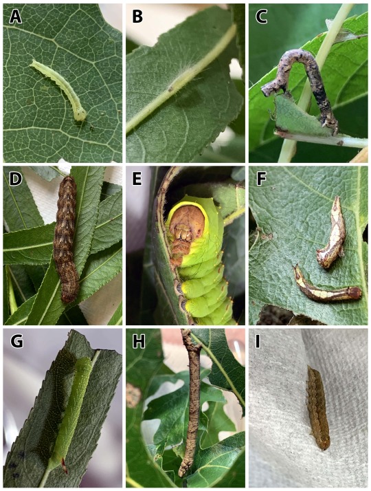
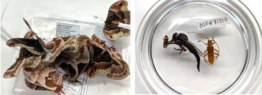
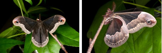
In 2018 a culture of Callosamia promethea caterpillars were reared.  A record of each stage of metamorphosis was preserved in alcohol and stored with reared caterpillars in the collection. Some of the cocoons were kept alive to allow the adult moths to emerge.  The adults were then preserved in the freezer so they could be prepared and added to the collection as a record of the offspring from that culture.

moths on leaves at night

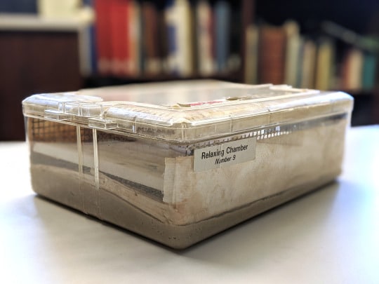
The moths were placed in a humidifying chamber overnight.  This chamber is kept humid by adding water to the absorbent paper towels that are layered in above a layer of sand mixed with an antifungal agent that keeps the specimens from getting moldy.

container labeled moth relaxing chamber number 3

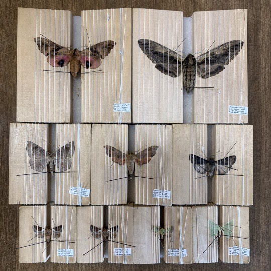
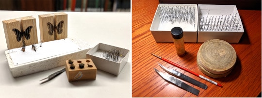
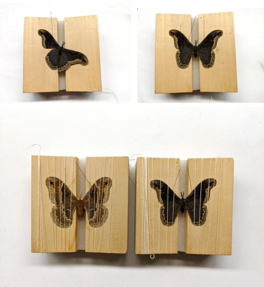
The specimens were thawed, and moisture kept the specimens relaxed enough to handle.  Specimens were pinned through the thorax and placed in a wooden pinning block designed for spreading the wings.

lepidoptera specimen laid out on blocks

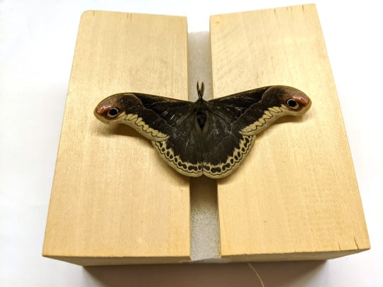
A series of very thin pins (size #000) were used to arrange the wings by carefully moving the forewings up high enough to expose the hindwings.  String was then wrapped around the block to hold the wings down and allow the specimens to dry.  Spread specimens remain on the spreading blocks for about a week to ensure they are completely dry and remain in the desired position.  Spreading moths and butterflies allows for all the characters on the hindwings to be visible, and it also allows the underside of the specimen to be viewed more easily.  The string is carefully unwrapped a week later, and specimens are removed from the blocks and ready to be labelled.

lepidoptera specimen on blocks in various stages of preparation

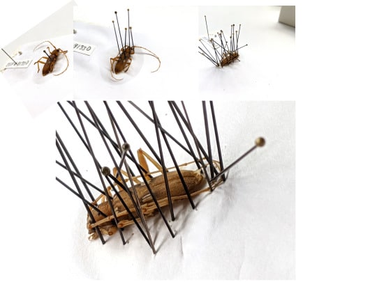
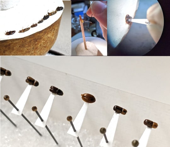
Non-Lepidoptera are usually pinned straight out of alcohol, when they are flexible enough to handle.  If they are collected and kept dry before preparation, then a relaxing chamber may be used to rehydrate them.  After the pin is inserted, the specimen is placed on a Styrofoam board lined with white paper.  Legs, antennae, and wings are arranged using brace pins that hold everything close to the body.  Specimens remain on the board for about a week until they are fully dry.

pinned specimens

If specimens are too small to be pinned, they are mounted on paper points using shellac glue.  The pin goes through the point made from archival paper using a tool known as a point punch.  After laying the specimens with the underside facing up, the tip of the point gets a dab of glue and each specimen is glued on to the tip of the point.

specimens mounted on paper

Guess what the next step is…labeling!

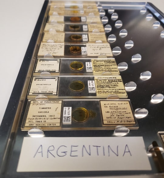
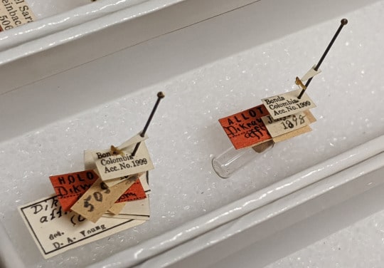
As mentioned before, specimens tell an important story.  The data is just as valuable as the specimens, and that data is printed on archival paper using a laser printer.  A labeling block is used to apply the label on the pin, below the specimen.  Any specimen that is prepared and labeled is ready to be identified and curated into the collection.

materials for labeling specimens

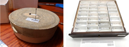
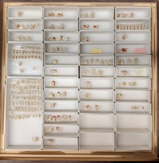
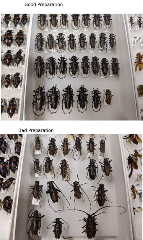
In a collection with 13.5 million specimens, space is valuable.  Well prepared specimens take up less space and are less vulnerable to damage.  A damaged specimen is still valuable because of the story it tells through its labels, even if damage makes the story incomplete.  High-quality preparation is important because it allows for easier examination of the specimens and interpretation of the differences between species.  These specimens are not just a bunch of bugs—all together, they are part of the record of life on Earth.

examples of good specimen preparation and bad specimen preparation

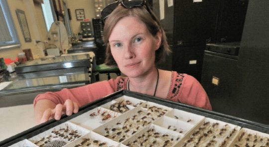
Vanessa Verdecia is Scientific Preparator in the museum’s Invertebrate Zoology Section. Museum employees are encouraged to blog about their unique experiences and knowledge gained from working at the museum.

Related Content

Traub Flea Data Books

Hillbilly Gourmets: Snail-eaters of the Yew Mountains

Ask a Scientist: What is a Caterpillar Database?

Filed Under: Blog Tagged With: Invertebrate Zoology, Science News, Section of Invertebrate Zoology, SWK2, Vanessa Verdecia

August 17, 2020 by wpengine

Traub Flea Data Books

With work-from-home restrictions in place, I’ve been transcribing the handwritten field notes (Figures 1-2) of world-renowned flea expert Robert Traub into a digital database. Between 1995 and 1997, Traub donated most of his collection to CMNH. Materials housed in the Traub collection span the globe, from the middle east to central America to islands in the pacific and beyond. The notebook I’m currently transcribing dates back to the mid-1900s, with records from particular field expeditions to Pakistan and Mexico.

Figure 1. Some of the notebooks written by Robert Traub containing information on his flea specimens.
Figure 2. One of the pages from a Traub notebook illustrating the specimen data it contains. Eventually, all of this data will be transcribed into electronic format so it can be searched and shared.

This type of retroactive data capture allows us to put standard locality information on specimens formerly associated with just an identification or data code number. This process also allows us to verify and update taxonomic names as necessary. While it’s not nearly as fun as field work, data capture and transcribing are still an important part of collections work.

The Traub collection is estimated to contain nearly 75,000 specimens mounted on glass slides (Figure 3), with 5,000 associated genitalic dissections. The enormous collection is housed in antique cabinetry as well as modern Eberbach cabinets. Almost 7,000 of these specimens only have a data code; thus, my digitization efforts and subsequent labeling continue!

Figure 3. Several slides from the Traub Flea collection.

Since I started working in IZ nearly three years ago, I have had the distinct privilege of working with different taxa every few months. From Lepidoptera, to Odonata, to Coleoptera, to Arachnida, and now Siphonaptera, these tasks serve as beautiful reminders of the diversity of life here on planet Earth.

Catherine Giles is Curatorial Assistant in the Section of Invertebrate Zoology at Carnegie Museum of Natural History. Museum employees are encouraged to blog about their unique experiences and knowledge gained from working at the museum.

Related Content

Moth Meme Science

Working with the Type Collection

A Bumble’s Blog and Bumble “Weee” Catapult Craft

Filed Under: Blog Tagged With: Catherine Giles, Invertebrate Zoology, Museum from Home, Science News, Section of Invertebrate Zoology

August 12, 2020 by wpengine

Working with the Type Collection

The staff of the Section of Invertebrate Zoology are currently working on many projects.  One of those projects is gathering, organizing, and taking inventory of our type collection materials.

Image 1.  A drawer of Lepidoptera types from the Invertebrate Zoology collection at CMNH.

Type specimens are the specimens upon which the scientific name and description of a species are based.  In other words, when scientists describe a new species, they use particular specimens to characterize the unique features of that particular species. Once a new species description is published, the specimen(s) used in the process of formally naming and describing the species become the type specimens.

Image 2. The original description of Dikraneura affinis (a leafhopper) as published in the Annals of Carnegie Museum, Vol. 18.

As such, type specimens are very valuable to science. Museum type collections serve as a sort of ‘library’ of species for scientific researchers.  With millions of insect species found on the earth, identification of a specimen you have in hand can be a daunting task.  Researchers around the world can compare specimens in their own collections with these scientific types to apply species-level determinations.

Image 3.  The plates included as part of the original species description.  Characteristics of Dikraneura affinis are illustrated in Figs. 24, 24a, and 24b.

It is estimated that there are approximately 40,000+ type specimens in the Invertebrate Zoology collection at CMNH. A more complete inventory and cataloguing of the type specimens in Invertebrate Zoology is currently required so that our type collections can be of greater use to the scientific community.

Image 4.  A drawer containing Hemipteran types, including Dikraneura affinis.  These particular insects are very tiny and are adhered to paper points for preservation and study.
Image 5.  Tray containing the two type specimens of Dikraneura affinis and their associated labels.

Prior to the availability of computers, George Wallace, a curator who worked at the museum on Hymenoptera from the 1930s to the 1970s, compiled and maintained a card catalog file of non-lepidopteran types in the Invertebrate Zoology collection.

Image 6.  Image of ‘card catalog’ of types compiled and maintained by curator George Wallace.
Image 7.  A sampling of the cards and associated information captured by George Wallace.

Many of the cards contain information about the published species descriptions, the numbers and kinds of type specimens in our collection, and label data associated with the specimens, including geographical, accession number, date, and collector information.

Image 8.  Type card showing information for Dikraneura affinis (a leafhopper).

To assist in the type collection organization effort, I have been tasked with digitizing over 1100 of these cards.  Digitizing the information renders it searchable and accessible to staff and allows for a more accurate inventory of our types.

Image 9.  Screenshot of file containing digitized data from Wallace type cards.  Information for Dikraneura affinis is highlighted on the screen.

Currently, researchers must contact us directly with queries that relate to our type collection. In the future, we hope to photograph our type specimens and make all of their specimen data available via the internet so that researchers worldwide may have access to the invaluable type collection resource that resides in the Section of Invertebrate Zoology at CMNH.

Hillary Fetzner is a Laboratory Assistant in the Section of Invertebrate Zoology at Carnegie Museum of Natural History. Museum employees are encouraged to blog about their unique experiences and knowledge gained from working at the museum.

Related Content

Delving into Lepidoptera Life History Studies

Meet Ainsley Seago, New Assistant Curator of Invertebrate Zoology

Ask a Scientist: Why do moths love lamps? 

Filed Under: Blog Tagged With: Hillary Fetzner, Holotypes, Invertebrate Zoology, Science News, Section of Invertebrate Zoology

August 11, 2020 by wpengine

Delving into Lepidoptera Life History Studies

For a number of years now in the Section of Invertebrate Zoology (IZ), we have been rearing larvae (= caterpillars) of different species of Lepidoptera (moths & butterflies) for both fun and research. This summer, given the ongoing COVID-19 pandemic and the need for everyone to isolate, I have taken to collecting and rearing a number of different species at home that were collected at a bug sheet in my own back yard (Figure 1).

Figure 1. A set up “bug sheet” used to attract insects at night.

Female moths are collected live and held in a plastic jar we call a “live jar” (Figure 2), until they lay eggs. If eggs are laid, and they are fertile, they usually hatch in about 7-10 days. This gives you enough time to identify the female adult to species (a recent field guide to moths and butterflies is a good place to start) so you can find out information on its preferred food source(s), or host plant(s), before the little larvae hatch are start searching around for food. If the eggs do hatch, rearing the resulting little caterpillars is a fun way to break up the tedium of being cooped-up at home for so long and is a nice way to bring Nature indoors.

Figure 2. Plastic “live jar” used to hold female moths until they lay eggs for rearing.

We have a little bit of experience rearing caterpillars at home. As you may know from a previous IZ blog post, my son and I reared some caterpillars that were not yet known to science, which resulted in a small publication. Right now, we have caterpillars of ten different species at various developmental stages. I check on them daily, making sure to keep their containers clean, and provide them with enough food to eat from their preferred host plant (Figure 3). It is amazing how quickly these little guys grow and change, all in the matter of a few short weeks. I try to capture images of them as they develop (see Figure 4), so they can be used on our websites, in blog posts (such as this one), or in eventual scientific publications that may result from the work.

Figure 3. Clear plastic rearing chambers containing caterpillar cultures, each started from eggs laid by a single female moth (= iso-female culture). Host plants include Maple, Willow, Oak, Sassafras, Cherry and Poplar.
Figure 4. Images of various species of caterpillars currently being reared by the author at home. A.) Early instar of Gluphisia septentrionis (Notodontidae), B.) Early instar of Acronicta dactylina (Noctuidae), C.) Later instar of Metarranthis sp. (Geometridae), D.) Last instar of Lithophane disposita (Noctuidae), E.) Later instar of Antheraea polyphemus (Saturniidae), F.) Two different early instars of Heterocampa obliqua (Notodontidae), G.) Early instar of Paonias excaecata (Sphingidae), H.) Last instar of Besma quercivoraria (Geometridae), I.) Later instar of an unknown caterpillar that was found on host plant food obtained for other caterpillars. The species will be determined when the adult moth emerges from the pupa later in the summer.

Once the females have laid eggs, they usually die as a result, having completed their task in the moth’s life cycle.  The females are then pinned, and the wings are usually spread on wooden blocks until they dry, so that the specimens can be easily identified and examined by experts in the future (Figure 5).  They then receive data labels that includes information on the specific locality and date of collection, method of collection, and the collector name(s).

Figure 5. Moths that have been pinned with their wings spread to aid in identification. Note the data labels have been associated with each specimen (lower right of each block).

My son and I are looking forward to watching our little menagerie of caterpillars progress throughout the summer, eventually completing their life cycle and becoming adult moths. I’m glad that we are able to give you a glimpse of our progress to date and hope you have enjoyed seeing some of these diverse little spineless wonders. Hopefully, when we can all return to our normal outdoor activities, you will have a newfound appreciation for these amazing insects when you encounter them out in the wild.

James W. Fetzner Jr. is Assistant Curator of Invertebrate Zoology at Carnegie Museum of Natural History. Museum staff, volunteers, and interns are encouraged to blog about their unique experiences and knowledge gained from working at the museum.

Related Content

Ask a Scientist: What are Murder Hornets?

Bug Bonanza: Virtual Bug Display

Is This Butterfly Blue or Green?

Filed Under: Blog Tagged With: Invertebrate Zoology, James Fetzner, Museum from Home, Science News, Section of Invertebrate Zoology

August 10, 2020 by wpengine

Meet Ainsley Seago, New Associate Curator of Invertebrate Zoology

Dr. Ainsley Seago (pictured with Vespula germanica, one of Australia’s many, many invasive species). Photograph by Jude Keogh.

Dr. Ainsley Seago studies the evolutionary history of beetles, from systematics and diversification of Staphylinoidea (rove, carrion, and fungus beetles) to the evolution of iridescence in Curculionoidea (weevils and their relatives). She has used everything from rotting squid traps to synchrotron radiation to better understand beetles in all their glory, but believes that the most important tool of all is a strong museum collection. Dr. Seago is thrilled to work with the CMNH collection and exhibit teams to bring the museum’s outstanding invertebrate collection to a wider audience, while using it to support research in Pittsburgh, the US, and beyond.

Dr. Seago is originally from Tacoma, WA,  and has just returned to the U.S. after 12 years in Australia.

Abstract of recent research (bearing in mind that I have a very loose grasp on what 8th graders are up to these days)

Australian stag beetle, Lamprima aurata (Coleoptera: Lucanidae). Photograph by Lauren Drysdale.

Among the world’s beetle species are hundreds of “living jewels,” insects with stunning jewel-like colors or shining golden armor. These so-called structural colors arise from nanoscale patterns in the exoskeleton, from variations in the thickness of chitin layers to intricate three-dimensional crystal lattices. Because they’re made by fixed structures and not chemical pigments, these types of insect color last indefinitely– even through fossilization.

Within the last 20 years, scientists have learned that several species of weevils (not to mention butterflies and longhorn beetles)  make their glittering, sequin-like colors with microscopic lattices called three-dimensional photonic crystals. We have also learned that these photonic crystals can generate different colors depending on how tightly spaced they are. However, the evolutionary origins of this type of iridescence have never been explored.

Iridescent scales of Pachyrhynchus orbifer (Coleoptera: Curculionidae). Photograph by Ainsley Seago.

Working with researchers from Yale-NUS (Singapore) and the Australian National Insect Collection (Canberra), I have conducted the first ever research placing a wide variety of photonic crystal structures from across the weevil family tree in an evolutionary (“phylogenetic”) context. The surprising result was that these crystals, found in hundreds of species of weevils, all derive from a single ancestral origin. Although three-dimensional photonic crystals have evolved repeatedly in insects, they appear to have evolved only once in weevils. The weevil lineages that gained these iridescent crystals then diversified rapidly, suggesting that the jewel-like colors aren’t just beautiful, they also confer a distinct evolutionary advantage.

Related Content

Meet Lisa Haney, New Postdoctoral Assistant Curator of Egypt on the Nile

Meet Amanda Martin, New Postdoctoral Research in the Section of Amphibians and Reptiles

Bug Bonanza: Jumping Spiders

Filed Under: Blog Tagged With: Ainsley Seago, Museum from Home, Science News, Section of Invertebrate Zoology

August 7, 2020 by wpengine

Mobilizing Millions of Marine Mollusks: Seashells by the Eastern U.S. Seashore

“What a pretty seashell, where did it come from?”

Perhaps the most important information that natural history museums keep about their specimens is where they came from. For many researchers, locality information is more important than the specimen itself. The specimen is useful to verify correct identification, but you can’t look at a specimen to determine where it came from.

As more and more museums share their specimen databases on-line, locality information is being used to document changes in distributions of organisms, including new occurrences of invading species, range shifts due to climate warming, and the disappearance of species becoming locally extinct.

Pretty seashells.

To facilitate uses of locality information, museums are scrambling to georeference their specimens. This term refers to the electronic pairing of the historic recorded location for each collected specimen with an established system of latitude and longitude coordinates. Georeferencing can be tedious and time-consuming, what with interpreting messy handwriting, dealing with misspellings, and tracking down obscure names, some of which have changed over time. Once I spent over an hour and got only two specimens georeferenced.

The US National Science Foundation (NSF) recognizes the importance of georeferenced specimens to facilitate understanding of where species occur and how their distributions change over time. Consequently, NSF has awarded $58,762 to Carnegie Museum of Natural History as one of 14 collaborating museums on a $2.3 million grant for a project titled: Mobilizing Millions of Marine Mollusks of the Eastern Seaboard. The project is spearheaded by Rudiger Bieler at The Field Museum in Chicago.

The main goal of the project is to georeference, and make available online, 535,000 lots representing 4.5 million specimens of marine mollusks (snails, clams, etc.) from the eastern USA. Only 15% of Eastern Seaboard mollusks in museums are currently reliably georeferenced. To facilitate georeferencing and promote standardization, each collaborating museum will focus on georeferencing all lots from particular geographical areas. Notably, for the first time, these museum records will distinguish between live- and dead-collected specimens, important information given that shells of dead mollusks sometimes persist for hundreds of thousands of years, and can be moved by currents and other animals such as hermit crabs. Whether or not a shell was collected alive is therefore crucial information for studies of biotic change using mollusks.

Two lots of East Coast USA seashells ready to georeference.

For CMNH, this award primarily means support for georeferencing our 11,436 lots of marine mollusks from eastern USA. In addition, we will catalog the eastern US part of our backlog, image relevant type specimens, create an exhibit, and, the aspect I am most excited about is creating an IPT, or integrated publishing toolkit, which will allow automatic updates from our in-house database to our web presence in the InvertEBase Symbiota portal.

The grant-funded new public display will interpret our biologically, commercially, and recreationally important marine mollusks from the Eastern Seaboard, and showcase mollusk diversity. The display will appeal to anyone who has beachcombed shells. Labels will describe how scientists use modern and historical specimens to study change in marine ecosystems over time. My hope is that visitors will learn that mollusks are diverse and beautiful, that museum collections are useful, and that evidence-based studies show ecosystem changes.

The Eastern Seaboard region includes 18 states, nearly 6,000 km of coastline, and about 3,000 molluscan species. Boundaries, from Maine to Texas, stretch from the shore outward to the edge of the U.S. Exclusive Economic Zone. The 14 collaborating U.S. collections contain 85% of all Eastern Seaboard marine mollusk museum holdings. These museum holdings average 8 specimens per lot – a lot is one species from one place at one time.

One hundred million mollusk specimens have been documented in natural history collections across North America. Each mollusk species in these collections average 1100 individuals, revealing geographic and morphological variation, and making mollusks among the best sampled group of metazoans, or multi-cellular animals. So far, freshwater and terrestrial mollusks have dominated digitization efforts of mollusks. This project is the first to focus on marine mollusks.

Shells are bio-archives. Shell skeletons record information about the animal and its environmental conditions throughout its life cycle. Shell material can be used to infer past ocean temperatures, seasonal fluctuations, and growth rates. Shell testing can reveal presence of trace elements and pesticides, allowing detection and identification of marine contamination and pollution.

In addition to their use in documenting what lived where and when, mollusks are important in other ways. Shells bring us joy when we find them on the beach. And many of us eat them. In 2016, three of the top 10 most valuable fisheries in the US, worth hundreds of millions of dollars, were mollusks: scallops, clams, and oysters.

Timothy A. Pearce, PhD, is the head of the mollusks section at Carnegie Museum of Natural History. Museum employees are encouraged to blog about their unique experiences and knowledge gained from working at the museum.

Related Content

Ask a Scientist: What is the biggest snail?

The Zebra Mussel and the Shopping Cart

Octopus Mystery: How Do They See Color?

Filed Under: Blog Tagged With: Museum from Home, Science News, Section of Mollusks, Tim Pearce

  • « Go to Previous Page
  • Page 1
  • Interim pages omitted …
  • Page 30
  • Page 31
  • Page 32
  • Page 33
  • Page 34
  • Interim pages omitted …
  • Page 43
  • Go to Next Page »

sidebar

About

  • Mission & Commitments
  • Directors Team
  • Museum History

Get Involved

  • Volunteer
  • Membership
  • Carnegie Discoverers
  • Donate
  • Employment
  • Events

Bring a Group

  • Groups of 10 or More
  • Birthday Parties at the Museum
  • Field Trips

Powdermill

  • Powdermill Nature Reserve
  • Powdermill Field Trips
  • Powdermill Staff
  • Research at Powdermill

More Information

  • Image Permission Requests
  • Science Stories
  • Accessibility
  • Shopping Cart
  • Contact
  • Visitor Policies
One of the Four Carnegie Museums | © Carnegie Institute | Privacy Policy | Terms of Use | Accessibility
Rad works here logo