• Skip to primary navigation
  • Skip to main content
  • Skip to primary sidebar

Carnegie Museum of Natural History

One of the Four Carnegie Museums of Pittsburgh

  • Visit
    • Buy Tickets
    • Visitor Information
    • Exhibitions
    • Events
    • Dining at the Museum
    • Celebrate at the Museum
    • Powdermill Nature Reserve
    • Event Venue Rental
    • Gift Cards
  • Learn
    • Field Trips
    • Educator Information
    • Programs at the Museum
    • Bring the Museum to You
    • Guided Programs FAQ
    • Programs Online
    • Climate and Rural Systems Partnership
  • Research
    • Scientific Sections
    • Science Stories
    • Science Videos
    • Senior Science & Research Staff
    • Museum Library
    • Science Seminars
    • Scientific Publications
    • Specimen and Artifact Identification
  • About
    • Mission & Commitments
    • Directors Team
    • Museum History
  • Tickets
  • Give
  • Shop

Enjoy the Museum from Home via our Blog

Can't make it to the museum in person? We've done our best to help cultivate resources for you to enjoy from home. Activities for the whole family, different ways to experience our exhibitions and more are included in these blogs.

November 6, 2020 by wpengine

Why Do Leaves Change Color?

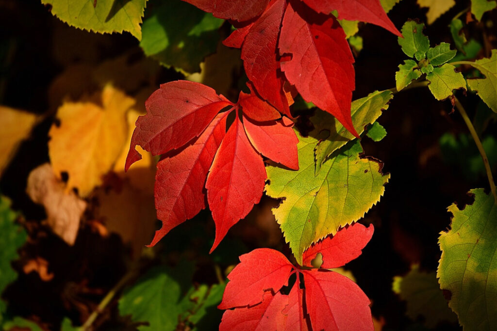
Image by 👀 Mabel Amber, who will one day from Pixabay.

This fall seemed to sneak up on me as time has been simultaneously moving at a rapid pace and in slow motion. As someone once told me when I became a parent, “the days are long, but the years are short,” and this global pandemic brings those words even closer to home. Nevertheless, I took the time to slow down and bask in the beauty of the changing fall leaves this October.  For me, nature is truly restorative, and there aren’t many things more beautiful than driving through the mountains of Western Pennsylvania during peak color change.

But, why do leaves change their color?

During the warmer months in Pennsylvania, trees take advantage of the increasing amount of light and good weather available from longer and warmer days. Using their leaves, trees absorb energy from sunlight, breathe in carbon dioxide, drink up water to produce their own food sources – sugar and starch. This process is only possible through chlorophyll housed in the leaf cells – giving leaves their vibrant green coloration.

Leaves also contain other color pigments ranging from yellow to orange – these pigments are often masked by great amounts of green coloring. But in the fall, the tree begins to prepare for shorter days and colder weather, and the leaves stop their food-making process. To prepare for the upcoming winter, the chlorophyll begins to break down, causing the green color to disappear. This change allows the ever-present yellow, orange, and red pigments to become visible. While chlorophyll breaks down, other chemical changes in the leaves can occur, creating an additional ray of colors through the development of red anthocyanin pigments. The yellow and orange pigments mixed with the red anthocyanin pigments give rise to the reddish and purplish fall colors of trees, such as dogwoods and sumacs.

Each species of tree shows off their own fall color. All these colors are due to the mixing of varying amounts of the chlorophyll residue and other pigments in the leaf during the fall season. This winter preparation creates a nature show like no other.

Another aspect of the leaves changing colors is that those leaves will eventually drop to the ground. Most of the broad-leaved trees in Pennsylvania shed their leaves in the fall (some trees retain their dead leaves until new growth starts in the spring). So, what should you do with your leaves in the fall? According to the U.S. Environmental Protection Agency, leaves and other yard debris account for more than 13% of the nation’s solid waste—33 million tons a year. In typical landfills, there isn’t enough oxygen to decompose the yard waste, causing the development and release of the greenhouse gas methane.

So, what do I do? LEAF IT! (get it?). Leave your leaves where they fall in the fall – or at least, find a nice place in your yard to pile the leaves. Leaf litter can act as both a fertilizer returning the nutrients back into the ground, and as a weed suppressant by acting as a ground cover. Leaf litter is also a vital habitat for much of our favorite wildlife. Many critters – from insects to mammals and everything in between – rely on leaf litter for food, shelter, and nesting material. Many of our favorite moth and butterfly caterpillars overwinter in fallen leaves before emerging in spring! So, if you want free mulch and fertilizer, to create wildlife habitat, and have more free time – LEAF IT!

Heather Hulton VanTassel is Assistant Director of Science and Research at Carnegie Museum of Natural History. Museum employees are encouraged to blog about their unique experiences and knowledge gained from working at the museum.
Related Content

Ask a Scientist: What makes poinsettias so special?

Turkeys

Clams in the Concrete! How Old is This Sidewalk?

Filed Under: Blog Tagged With: Botany, Heather Hulton VanTassel, Museum from Home, Science News

November 6, 2020 by wpengine

The 12,000-Year Journey Of The Cheeseburger

In one large bite, a bun, ground beef patty, cheese, lettuce, and tomato could finally fulfill its purpose: to be my lunch. Many people have seen ads for, or even eaten a cheeseburger before. But where do all the ingredients come from? The tasty combination of meat, vegetables, grain, and milk product has 12,000-year-old roots in a faraway land across the sea. From there, over thousands of years and thousands of miles, it made a journey to its ultimate destination … my stomach. As delicious as it is, every good ending has a story.

The Bun

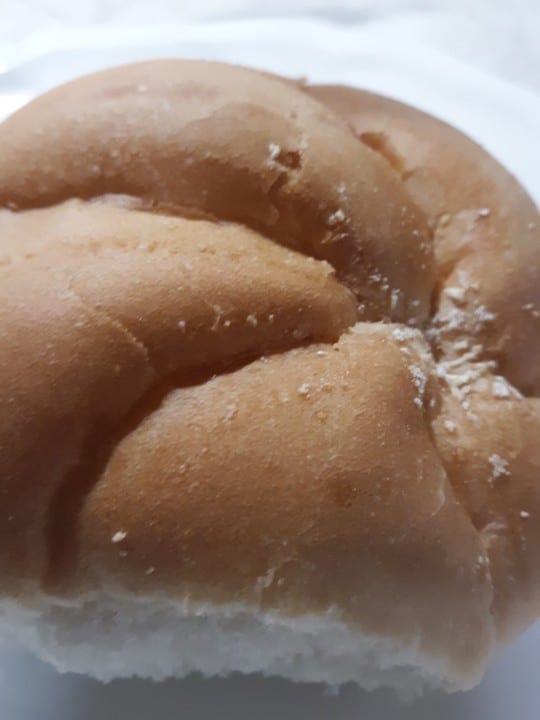
For a proper burger, you need the bun to sandwich all its deliciousness. The main ingredient for the bun is flour, which comes from wheat. Today, there are 25,000 distinct forms of wheat, all descended from a plant called emmer, which first originated in the Fertile Crescent within the Middle East. The earliest evidence for emmer being deliberately grown by humans for food (domestication) was from at least 12,000 years ago.

Ancient humans, just like us today, enjoyed eating wheat products (I love my pizza!). Where it grows abundantly, wheat is easily harvested and can be stored for extended periods of time, making it a stable source of vegetable protein. Thus, some of the first civilizations, like the Babylonians and Assyrians, sprung up in the Fertile Crescent. Emmer wheat spread to Greece, Cyprus, and India by 6500 BCE, and to Egypt shortly after. In fact, the Egyptians are the first people known to make bread.

close up of hamburger bun

The Patty

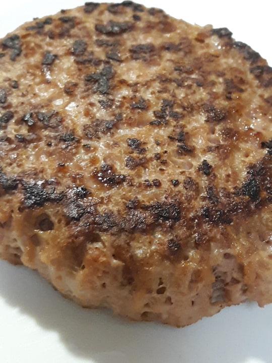
Now let’s get to the deliciousness housed between the buns: the patty. Traditional cheeseburgers are made from beef, which comes from cattle. Unlike emmer wheat, cattle, which descended from wild oxen called aurochs, were domesticated separately in two (possibly three) different places: the Fertile Crescent, the Indus Valley (modern-day Pakistan), and possibly northeast Africa 10,000-8000 years ago. From there, domesticated cattle spread across the continents of Africa, Asia, and Europe.

Cattle were one of the first mammals to be domesticated. They provide many useful products used for consumption (meat, milk, fat) and tool making (horns, hooves, hides). Additionally, their large size allowed them to pull heavy objects like plows for farming. Because of their importance, many religions and cultures considered cattle to be sacred. In Ancient Egypt, many of their gods had cattle forms, including Hathor, Ptah, Menthu, and Atum-Ra, Ancient Greeks often used cattle as sacrifices to the gods. Even today, Hindus do not eat cattle meat.

close up of burger patty

The Cheese

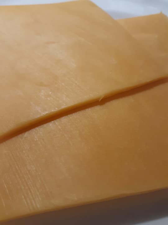
Finally, a cheeseburger would hardly be a cheeseburger without the cheese (which is made from milk). Although cow milk is the most popular source material today, cheese was originally made from goat or sheep milk. Cheesemaking began over 4,000 years ago, but how it started is unclear. Legend has it that it was an Arabian merchant who accidentally created the first cheese. He put his milk in a pouch made from a sheep’s stomach as he traversed across the desert. Sheep stomachs contain an enzyme called rennet, and when the milk chemically reacted to the enzyme and heat from the sun, it separated into curd and whey. The curd is what we commonly refer to as the cheese.

Although cheesemaking’s origins remain ambiguous, the Romans were the first to make cheesemaking a widespread industry. Aging and smoking cheese extends the product’s shelf-life, enabling Roman soldiers to carry this excellent source of protein with them. As they conquered the European continent, they spread their cheesemaking. At the height of the Roman empire, they were making and trading hundreds of different kinds of cheese. Only later during European colonization was cheese spread to the Americas and Asia.

slices of yellow cheese

The Cheeseburger

So what genius put it all together? None other than a 16-year-old named Lionel Sternberger. His father owned a sandwich shop, and one day in 1924, Lionel put a slice of American cheese on one of his father’s hamburgers. He called it a “cheese hamburger.” One decade later, a Kaelin’s restaurant in Louisville, Kentucky gave the sandwich the name “cheeseburger,” which was trademarked in 1935 by Louis Ballast of Humpty Dumpty Drive-In.

man wearing pink glasses and a hat holding a cheeseburger

The End (of This Story of Deliciousness)

Who knew that there was so much behind a basic cheeseburger? From sheep stomach pouches to Babylonians, each played a role in creating the cheeseburger in your hands. Even Pittsburgh has some cheeseburger fame! Did you know that Jim Delligatti, who owned a restaurant in Uniontown PA, part of the Greater Pittsburgh Region, created the McDonald’s Big Mac in 1967?

Angela Wu is a Teen Volunteer in the Education Department. Museum employees, volunteers, and interns are encouraged to blog about their unique experiences and knowledge gained from working at the museum.
Sources:

The Big Mac turns 40, gets a museum. (2007, August 26). ABC News. Retrieved August 9, 2020, from https://abcnews.go.com/Business/story?id=3524528&page=1#:~:text=The%20Big%20Mac%20was%20first,staple%20of%20McDonald’s%20menus%20nationwide.

Cooper, R. (2015, July). Re-discovering ancient wheat varieties as functional foods. ScienceDirect. Retrieved August 5, 2020, from https://www.sciencedirect.com/science/article/pii/S2225411015000401

Cownie, E. (2018, August 27). Why cattle mattered in the Ancient World. Medium. Retrieved August 8, 2020, from https://medium.com/@emmafcownie/why-cattle-mattered-in-the-ancient-world-4e27b1c37e58

Hirst, K. (2019, July 9). Wheat Domestication. ThoughtCo. Retrieved August 6, 2020, from https://www.thoughtco.com/wheat-domestication-the-history-170669

History of Cheese. (2020, January 25). International Dairy Foods Association. Retrieved August 6, 2020, from https://www.idfa.org/history-of-cheese

Mitzewich, J. (2020, May 15). Who Invented the All-American Cheeseburger? The Spruce Eats. Retrieved August 7, 2020, from https://www.thespruceeats.com/birth-of-the-cheeseburger-101426

Pitt, D., Sevane, N., Nicolazzi, E. L., MacHugh, D. E., Park, S., Colli, L., Martinez, R., Bruford, M. W., & Orozco-terWengel, P. (2018). Domestication of cattle: Two or three events?. Evolutionary applications, 12(1), 123–136. https://doi.org/10.1111/eva.12674

Roberts, B. (2018, March 5). The Fascinating 7,500 Year History of Cheese. Forbes. Retrieved August 5, 2020, from https://www.forbes.com/sites/brianroberts/2018/03/05/the-history-of-cheese/#4807da304ca1

Related Content

Potatoes, Sweet Potatoes, and Yams: What’s the Difference?

Hip and “Trashy” Ice Cream

Jurassic Days: Cookie Excavation

Filed Under: Blog Tagged With: Anthropocene Living Room, Museum from Home

November 4, 2020 by wpengine

The Bromacker Fossil Project Part XI: Dimetrodon teutonis, an apex predator

New to this series? Read The Bromacker Fossil Project Part I, Part II, Part III, Part IV, Part V, Part VI, Part VII, Part VIII, Part IX, and Part X. 

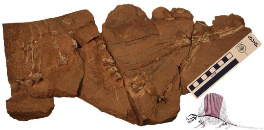
Holotype specimen of Dimetrodon teutonis, which consists of a partial vertebral column. The preserved portion of this vertebral column is highlighted in the reconstruction of Dimetrodon (lower right). Photograph by the author, 2007. Dimetrodon reconstruction modified from Romer and Price, 1940.

Specimens of two top predators have been discovered at the Bromacker quarry. Like Martensius, both are basal members of the group Synapsida, the later members of which gave rise to mammals. You might be familiar with one of them – Dimetrodon, a synapsid sometimes incorrectly portrayed with dinosaurs, which carried a tall sail on its back that was supported by bony spines. The other is a new genus and species that will be presented in my next post.

The fossil pictured above, the first-discovered specimen of Dimetrodon from the Bromacker quarry, may not look like much, but it was the first record of Dimetrodon outside of North America. The circumstances under which it was found were very different from the discovery of other fossils from the Bromacker quarry. Before Dave Berman and I arrived for the 1999 field season, Thomas Martens noticed that someone, possibly a fossil poacher, had been in the quarry overnight and knocked some rocks off the quarry lip. The rocks apparently broke upon hitting the ground, which exposed some bones. Thomas carefully picked them up and took them to his lab at the Museum der Natur, Gotha (MNG). When Dave and I met Thomas at the quarry on our first day of the field season, Thomas mentioned the find and told us that he thought the bones were ribs. We didn’t think much of it, other than horror at learning a fossil poacher might have visited the quarry overnight, one of our worst fears.

As planned, Dave and I spent the last day of the field season in the museum collections, and when Thomas let us in that morning, he reminded us to look at the potential ribs and told us where they were. Shortly after we began examining them, Dave and I simultaneously realized that the “ribs” were actually spines of Dimetrodon. We couldn’t believe our eyes, because of all the Early Permian fossils known from North America, Dimetrodon was Thomas’ favorite. Indeed, he’d used an image of it on signs at the Bromacker and included a model of Dimetrodon in a diorama, once on display in the MNG, that showed models of Bromacker animals in their environment. Thomas jumped for joy later that day when we gave him the news.

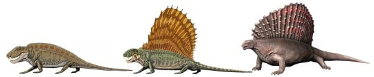
So how did Dave and I so quickly realize that the “ribs” were spines of Dimetrodon? Besides Dimetrodon, some other basal synapsids had sails, the function of which remains unknown, though scientists have speculated they could’ve been used for display or regulating body temperature. The spines (known as neural spines) supporting the sails vary in shape and length, with those of Dimetrodon and its herbivorous relative Edaphosaurus being tall and narrow, and those of another relative, the carnivorous Sphenacodon, being shorter and blade-like. Neural spines of Dimetrodon are easy to distinguish, because in addition to being long they bear fore and aft grooves, which create a dumbbell-shaped cross-sectional outline, and they lack the ‘crossbars’ that occur on the long neural spines of Edaphosaurus. When Dave and I saw the fore and aft grooves, the dumbbell-shaped cross-sectional outline of some broken spine ends, and an absence of crossbars, we knew that the “ribs” were indeed spines of Dimetrodon.

Flesh reconstructions of Sphenacodon sp. (left), Dimetrodon grandis (middle), and Edaphosaurus pogonias (right) to show the differences between their sails. Note that Dimetrodon and Sphenacodon are more closely related to one another than they are to Edaphosaurus, despite their different sail shapes. Reconstructions of Sphenacodon and Dimetrodon by Dmitry Bogdanov and that of Edaphosaurus by Nobu Tamura, all from Wikimedia Commons.

The Bromacker Dimetrodon is considerably smaller than other known species of the genus, and this is one character among other more detailed anatomical features that distinguishes it. For the new species name, Dave selected the Latin “teutonis,” which means an individual of a German tribe, in reference to the geographic origin of the holotype specimen.

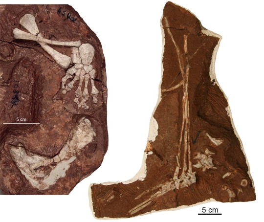
Two additional specimens of Dimetrodon teutonis. Left, hindleg and shoulder girdle bone (fused scapulocoracoid) and right, several vertebrae bearing complete to nearly complete neural spines of an individual that was larger and presumably more mature than the holotype. Photographs by the author, 2007.

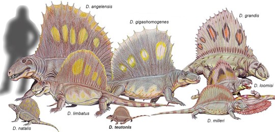
Dave was able to use a mathematical equation involving measurements of the vertebrae to estimate the holotype’s weight as a living animal at 31 pounds. In contrast, other known Dimetrodon species have estimated weights of about 81–550 pounds. We later discovered additional partial specimens of Dimetrodon at the Bromacker quarry, and Dave estimated the weight of the largest specimen with vertebrae at 53 pounds, still considerably less than that of what had previously been the smallest species, D. natalis from Texas. Dimetrodon is otherwise known from numerous species from the American mid-continent and southwest that generally got larger through time.

Reconstructions of various species of Dimetrodon drawn to scale. The diminutive D. teutonis is at bottom center and D. natalis, no longer the smallest species, is at bottom left. Illustration adapted from Dmitry Bogdanov via Wikimedia Commons.

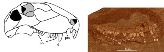
All Dimetrodon species have teeth adapted for meat-eating in being teardrop-shaped with sharp edges for slashing flesh. By size and jaw position these sharp teeth are divided into precanines, canines, and postcanines of varying numbers. Unlike D. teutonis, some species even had fine serrations on their tooth edges. The only known upper jaw bone of Dimetrodon teutonis clearly has two canines, but one is missing and represented by a large gap in the tooth row that would have accommodated this tooth. The second canine is represented only by its broad base, but it too must have been large. Although it was a small animal, the teeth of D. teutonis indicate that it was a meat-eater and as such would have preyed on other vertebrates from the Bromacker, many of which were even smaller.

Diagrammatic drawing of the skull of Dimetrodon (left) and photograph of the maxilla or upper jaw bone (right) of D.teutonis. Abbreviations: c, canine; pc, postcanine; prc, precanine. Photographs by the author, 2007. Drawing of skull from Wikimedia Commons.

Stay tuned for my next post, which will be about the second-known apex carnivore from the Bromacker. In the meantime, here are links to scientific papers on Dimetrodon teutonis:

https://www.researchgate.net/publication/325670232_A_new_species_of_Dimetrodon_Synapsida_Sphenacodontidae_from_the_Lower_Permian_of_Germany_records_first_occurrence_of_genus_outside_of_North_America

https://www.researchgate.net/publication/288544821_New_materials_of_Dimetrodon_teutonis_Synapsida_Sphenacodontidae_from_the_Lower_Permian_of_Germany

Amy Henrici is Collection Manager in the Section of Vertebrate Paleontology at Carnegie Museum of Natural History. Museum employees are encouraged to blog about their unique experiences and knowledge gained from working at the museum.

Keep Reading

The Bromacker Fossil Project Part XII: Tambacarnifex unguifalcatus, the Tambach Executioner 

Filed Under: Blog Tagged With: Amy Henrici, Museum from Home, Science News, Section of Vertebrate Paleontology, The Bromacker Fossil Project, Vertebrate Paleontology

October 6, 2020 by wpengine

Sympathy for the Devil

Bats and devils are among the most popular topics associated with Hallowe’en.  Of course, the research collection in the Section of Mammals has worldwide examples of bats species, but we don’t find them scary and we think about bats and their vital ecological roles all year long.  Perhaps more mysterious and less well-known are the two Devil specimens stored among the wombats, kangaroos, and koalas in our collection.  Even school children have heard about *our* kind of devils.  Yes, the Tasmanian devil (Sarcophilus harrisii) is a marsupial – a pouched mammal, like our opossum – that is found only on the island of Tasmania, located some 140 miles off the southeast coast of Australia.  Fossil evidence tells us that it once lived on the Australian mainland, but it may have been wiped out on the continent by the introduction of the Dingo, Australia’s legendary wild dog.

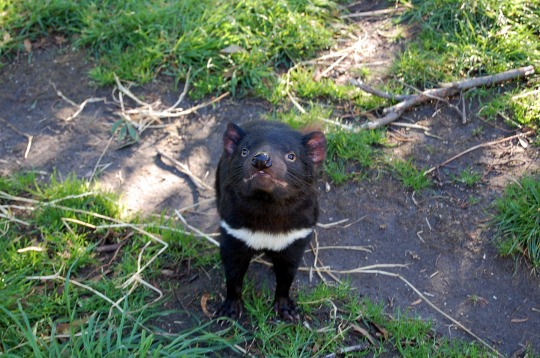
photograph of Tasmanian devil

The Tasmanian devil is a stocky mammal with short legs, short black fur and a distinctive white throat patch. Its head is noticeably large for the size of the body. An adult male may weigh up to 20 lbs. They are nocturnal with a good sense of sight, smell, and touch. Devils are known to cover significant distances nightly, in search of carrion or prey. They can move surprisingly fast and seem to enjoy swimming. In the wild, individuals can live between five and seven years, but many die within the first year of birth. Although it is the largest living marsupial carnivore, the Tasmanian devil is predominantly a scavenger.

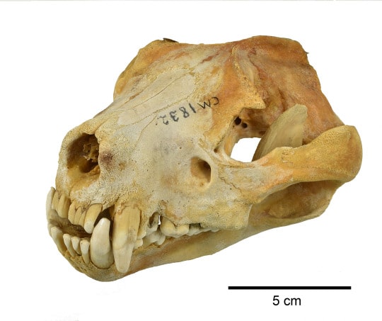
Tasmanian devil skull

A close look at the skull shows evidence of space on the side of the head for large jaw muscles. For its size, the Tasmanian devil has the strongest bite force of any mammal – more powerful than even a hyena! With the large masseter muscles and especially large molars, it can easily crush bone. In fact, devils are such efficient carrion-eaters that they willingly consume an entire carcass, including the fur.

Although this animal gained a reputation for having a bad disposition, it is speculated that this impression was derived from the poor conditions it was kept in when first captured for observations. Since then, it sometimes has been kept humanely as a pet and been found to be much friendlier than initially reported. Tasmanian devils do not seek each other’s company except during the mating period. However, they often come together to feed on a dead animal, where vocalizations and as many as nineteen different behavioral cues are used for communication. These communal gatherings are characterized by aggression and loud sounds, described as “frequent growling” and “blood-curdling screams”!

In 1996, a sad chapter began in the existence of the Tasmanian devil. A deadly infectious cancer called devil facial tumor disease, began to spread within the population. In 2012, the Australian government transferred 30 disease-free individuals to tiny Maria Island off the coast of Tasmania, in what was called ‘island insurance’, while researchers worked on perfecting a vaccine. By 2017, the disease had led to a 90% extinction rate on Tasmania. In hopeful news, by 2019 there were indications that surviving individuals’ immune systems may be undergoing modifications to fight the disease. In early September 2020, a consortium of conservation groups released 11 Tasmanian devils to a wildlife sanctuary in the state of New South Wales, placing the Tasmanian devil on the Australian mainland for the first time in more than 3000 years.  An additional 15 devils were released in early October and more releases are planned.

Currently, the Tasmanian devil is not extinct, but its recovery hangs in the balance. It would be tragic if we are left only with museum specimens and Taz, the Looney Tunes cartoon image, of this fascinating mammal.

Suzanne B. McLaren is the Collection Manager in the Section of Mammals at Carnegie Museum of Natural History. Museum employees are encouraged to share their unique experiences from working at the museum.

Related Content

Scientists Live: Mammalogist Suzanne McLaren on Bats

Vampire Squid: Cutest Dracula

Do Snakes Believe in the Tooth Fairy?

Filed Under: Blog Tagged With: mammals, Museum from Home, Science News, Section of Mammals, Suzanne McLaren

October 2, 2020 by wpengine

Vampire Squid: Cutest Dracula

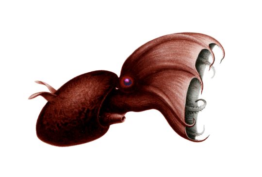
The Vampire Squid is your go-to mollusk for Halloween. It’s covered with glow-in-the-dark spots, and it can hoist its cape-like webbed arms over its head to transform into a pumpkin shape complete with outward-pointing fleshy spines. But wait, there’s more. With the largest eyes relative to body size of any animal, this has got to be the cutest Dracula you ever saw. And the scientific name, inspired by the cloak-like webbing and the dark body color, literally translates to “vampire squid from hell.”

Vampire Squid, showing cloak-like webbing between arms, large eye, and ear-like fins. [Image from Wikimedia Commons]

The Vampire Squid (Vampyroteuthis infernalis) is an extreme deep-water cephalopod more closely related to octopuses than to squids. It is so bizarre that scientists classify it in its own taxonomic order, Vampyromorphida, to show that it differs markedly from other living cephalopods. Like octopuses, it has 8 arms with webbing between them, but unlike octopuses that have suckers on the entire length of the arms, the Vampire Squid bears suckers only on their outermost half. The prominent feature on the arms of the Vampire Squid are fleshy spines or cirri. In addition to the eight arms, it has two velar filaments, in pouches in the webbing, that are analogous (and maybe homologous) to the two long tentacles of squids.

Regarding superlatives, the Vampire Squid has the largest eyes relative to its body size of any other animal, a detail noted in the Guinness World Records. A fully-grown individual can be 28 cm (11 inches) long with eyes 2.5 cm (1 inch) in diameter. Adding to the cuteness factor, they have adorable ear-like fins, which adults use for swimming; juveniles also have fins, but primarily use jet propulsion to move around.

They live in the lightless ocean depths 600-900 m (2000-3000 feet) deep in temperate and tropical oceans world-wide. The ocean at these depths is an oxygen minimum zone with so little dissolved oxygen that most complex organisms cannot survive. But the vampire squid survives perfectly well with a low metabolism and blue blood that is more efficient at carrying oxygen than that of other cephalopods. They use ammonium in their tissues to regulate their buoyancy (ammonium is a wee bit lighter than water), reducing the need for active swimming. Living in the oxygen minimum zone probably helps it to avoid predators.

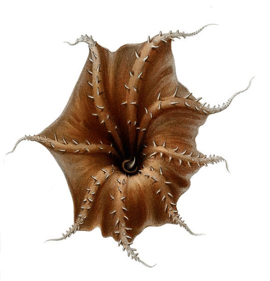
If disturbed, the Vampire Squid kind of turns itself inside-out into the “pumpkin” or “pineapple” posture by curling its arms and webbing up to cover the body with the spiny cirri pointing outward. Their body is covered by photophores, or light-emitting organs, which they can use to flash a wide range of patterns. In the pumpkin pose, they conceal most of the photophores, but they can light up the tips of the arms and wave them around to distract predators. If it gets really annoyed, the Vampire Squid can release a sticky cloud of luminous mucus that glows for nearly 10 minutes, presumably long enough for the Vampire Squid to make a get-away into the inky darkness.

Vampire Squid, underside of arms showing fleshy spines. [Image from Wikimedia Commons]

Much of what we know about their behavior comes from videos made by Remotely Operated Vehicles. It is hard to keep Vampire Squids alive in aquariums at the much lower pressure of our human world, but the Monterey Bay Aquarium succeeded for a while and has some great videos. Aquarium scientists were able to solve the mystery about what the Vampire Squid eats. No, it doesn’t eat blood! It eats detritus (organic debris), also known as marine snow. As the Vampire Squid drifts in the current, any debris that touches an extended filament is moved by the creature’s arms to its mouth. Unusual for being the only known cephalopod to eat non-living food, the Vampire Squid is adapted to eat material that falls through the oxygen minimum zone. Marine snow includes dead bodies, feces, and a lot of mucus from above, and because of the mucus, it is sometimes jokingly referred to as marine snot.

I imagine if Dracula learned about the Vampire Squid, he might exclaim, “I thought it was eating blood, but it’s snot!”

Timothy A. Pearce, PhD, is the head of the mollusks section at Carnegie Museum of Natural History. Museum employees are encouraged to blog about their unique experiences and knowledge gained from working at the museum.
Related Content

Extremely Rapid Evolution of Cone Snail Toxins

Ask a Scientist: Are slugs and snails the same thing?

Chasing Snails in the Great Smoky Mountains

Filed Under: Blog Tagged With: Museum from Home, Science News, Section of Mollusks, Tim Pearce

October 2, 2020 by wpengine

Do Snakes Believe in the Tooth Fairy?

When a child loses a baby tooth, the Tooth Fairy will sneakily appear a short time later to snatch that tooth up and leave behind a little treat. But what happens when vipers or other snakes with large fangs lose their teeth? I doubt the Tooth Fairy would be too keen about sneaking up on a sleeping snake…and as someone who studies and admires snakes, I would not recommend it!

image
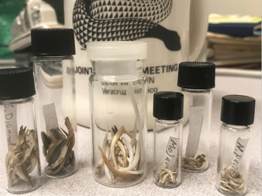
Vials containing Viperidae snake fangs. The middle vial (with clear lid) contains fangs of the Gaboon viper.

I recently learned the answer to this question when I was given seven tiny vials containing dozens of fangs. These fangs came from various species of snakes in the Viperidae family, including the Mojave green rattlesnake (Crotalus scutulatus), the Northern Pacific rattlesnake (Crotalus oreganus), and the Gaboon viper (Bitis gabonica), which has the longest fangs of any snake species. The researcher who gifted us these fangs was curious about the outcome of shed snake teeth and wanted to determine how frequently snake fangs may be swallowed and passed through the snake’s digestive tract. To answer this question, he dissected and examined dried snake feces for the possible presence of shed fangs.*

As it turns out, snakes will occasionally swallow their shed fangs! Vipers are carnivores that have to hunt down and subdue live prey in order to eat and survive. Often there is a struggle between predator and prey and, in that process, a fang may be wiggled lose. Instead of falling out of a snake’s mouth, the snake may swallow the fang along with the prey item. The fang will ultimately pass through the snake’s digestive system and emerge in its feces.

image
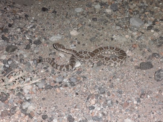
Pacific rattlesnake (Crotalus oreganus) in the Mojave Desert, California. One of the vials contained fangs from this species, but from snakes in the northern portion of the species’ range.

As the collection manager of the Section of Amphibians and Reptiles, I oversee and care for the museum’s massively beautiful and useful collection of cool and creepy herpetofaunal specimens. These specimens include full body wet specimens preserved in alcohol, the osteology collection of bones and turtle shells, and other items such as histology slides, gut contents, and even fangs. Regardless of their preservation form, all the reptile and amphibian specimens within the collection are useful for researchers and could serve to answer future scientific questions. Although the fangs within these seven seemingly unimposing vials have already answered one burning scientific question, they will be added to the collection so see what other answers they can provide!

*Researchers take careful precautions when handling feces as it can carry disease. Do not handle feces you may see in the wild.

Stevie Kennedy-Gold is the collection manager for the Section of Amphibians and Reptiles at Carnegie Museum of Natural History. Museum employees are encouraged to blog about their unique experiences and knowledge gained from working at the museum.
Related Content

Ask a Scientist: What is an Alcohol House?

The Enduring Appeal of Snakes

Poisons of the Carnegie: Timber Rattlesnake

Filed Under: Blog Tagged With: herpetology, Museum from Home, Science News, Stevie Kennedy-Gold

  • « Go to Previous Page
  • Page 1
  • Interim pages omitted …
  • Page 6
  • Page 7
  • Page 8
  • Page 9
  • Page 10
  • Interim pages omitted …
  • Page 26
  • Go to Next Page »

sidebar

About

  • Mission & Commitments
  • Directors Team
  • Museum History

Get Involved

  • Volunteer
  • Membership
  • Carnegie Discoverers
  • Donate
  • Employment
  • Events

Bring a Group

  • Groups of 10 or More
  • Birthday Parties at the Museum
  • Field Trips

Powdermill

  • Powdermill Nature Reserve
  • Powdermill Field Trips
  • Powdermill Staff
  • Research at Powdermill

More Information

  • Image Permission Requests
  • Science Stories
  • Accessibility
  • Shopping Cart
  • Contact
  • Visitor Policies
Stay in the loop! Sign up for our newsletter(s).
One of the Four Carnegie Museums | © Carnegie Institute | Privacy Policy | Terms of Use | Accessibility
Rad works here logo