• Skip to primary navigation
  • Skip to main content
  • Skip to primary sidebar

Carnegie Museum of Natural History

One of the Four Carnegie Museums of Pittsburgh

  • Visit
    • Buy Tickets
    • Visitor Information
    • Exhibitions
    • Events
    • Dining at the Museum
    • Celebrate at the Museum
    • Powdermill Nature Reserve
    • Event Venue Rental
    • Gift Cards
  • Learn
    • Field Trips
    • Educator Information
    • Programs at the Museum
    • Bring the Museum to You
    • Guided Programs FAQ
    • Programs Online
    • Climate and Rural Systems Partnership
  • Research
    • Scientific Sections
    • Science Stories
    • Science Videos
    • Senior Science & Research Staff
    • Museum Library
    • Science Seminars
    • Scientific Publications
    • Specimen and Artifact Identification
  • About
    • Mission & Commitments
    • Directors Team
    • Museum History
  • Tickets
  • Give
  • Shop

Blogs from our Scientific Researchers

Carnegie Museum of Natural History is home to active research and vast scientific collections. Our scientific researchers regularly contribute to the blog at the museum.

October 21, 2020 by wpengine

Collected on This Day 98 Years Ago

Chestnuts (used to be) on Chestnut Ridge

And across the entire state of Pennsylvania.
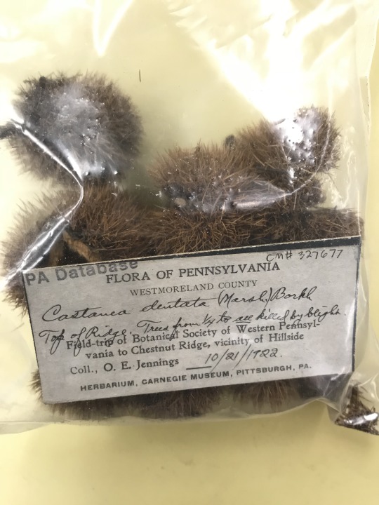
 

bag of chestnut seeds

American chestnut (Castanea dentata) was once a very common tree, native from Maine to Mississippi. In the heart of the Appalachians, the historical range covered the entire state of Pennsylvania. I say “historical” and “once a very common tree” because it is no longer.  You may occasionally stumble upon an American chestnut tree, especially small trees and saplings persisting as sprouts from the large trees that graced our landscape a century ago. Older trees, with mature fruits, are quite rare.  

In fact, some estimates suggest American chestnut accounted for one in four trees in some forests!  So, what happened?  In the early 1900s, a disease caused by a pathogenic fungus (Cryphonectria parasitica) was accidentally introduced with imported Asian trees. It was first recorded in New York City in 1904.  In a matter of decades, American chestnut was nearly decimated by this disease known as Chestnut blight.

The Carnegie Museum of Natural History herbarium captures this change in our forests.  
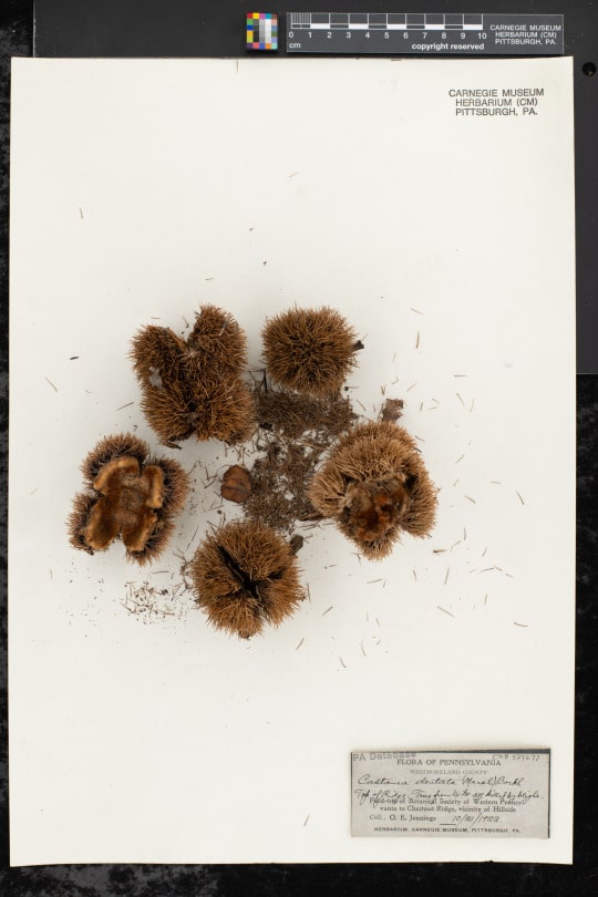
American chestnut specimen on herbarium sheet

This specimen of American chestnut was collected by influential Carnegie Museum curator Otto Jennings on October 21, 1922 on a field trip of the Botanical Society of Western Pennsylvania to Chestnut Ridge, near Derry Township, Pennsylvania.  Chestnut Ridge is a ridge of the Allegheny Mountains, presumably named for its (once) many American chestnuts.  

This specimen is from the fruit collection of the herbarium.  These specimens are different than the “standard” pressed flat specimens on paper.  Instead, they are stored to maintain their three-dimensional structure.

Note the note made by Jennings on the label on this specimen: “Trees from ¼ to all killed by blight.”

The case of the American chestnut is an interesting one.  It served important cultural and ecological roles; some even calling it a “keystone” species.  There is no doubt that the functional extinction of American chestnut ricocheted through the ecosystem, causing long-term biological changes. Many of these changes we may not know.  Yet, at the same time, despite the species importance, our forests continue.  Presumably other species have filled the functional and physical space of American chestnut.  

Disease and pest outbreaks in Pennsylvania’s forests continue.  Many of our critical tree species are likely to decline in coming years and decades.  Some iconic species have already declined or are at risk.  These include our ash species (mortality caused by introduced Emerald Ash Borer), American beech (Beech leaf disease, Beech bark disease caused by an introduced scale insect), and eastern hemlock (mortality caused by introduced sap sucking bug, the hemlock woolly adelgid)…to name only a few threats.

What will Penn’s woods look like in another 100 years?  

Our collections document the past and present to inform our decisions for the future.

Find this American chestnut specimen here (along with 268 others!): https://midatlanticherbaria.org/portal/collections/list.php?db=328&includecult=1&taxa=Castanea+dentata&usethes=1&taxontype=2

Check back for more! Botanists at the Carnegie Museum of Natural History share digital specimens from the herbarium on dates they were collected. They are in the midst of a three-year project to digitize nearly 190,000 plant specimens collected in the region, making images and other data publicly available online. This effort is part of the Mid-Atlantic Megalopolis Project (mamdigitization.org), a network of thirteen herbaria spanning the densely populated urban corridor from Washington, D.C. to New York City to achieve a greater understanding of our urban areas, including the unique industrial and environmental history of the greater Pittsburgh region. This project is made possible by the National Science Foundation under grant no. 1801022.

Mason Heberling is Assistant Curator of Botany at Carnegie Museum of Natural History. Museum employees are encouraged to blog about their unique experiences and knowledge gained from working at the museum.
Related Content

Our Eureka Moment! 

Super Science: Goldenrod

A Day Late, But Thanks for Your Impatiens! 

Filed Under: Blog Tagged With: Botany, collected on this day, Hall of Botany, Mason Heberling, Science News, Section of Botany

October 13, 2020 by wpengine

Is this what they call overkill? Toxin and venom in the herp world

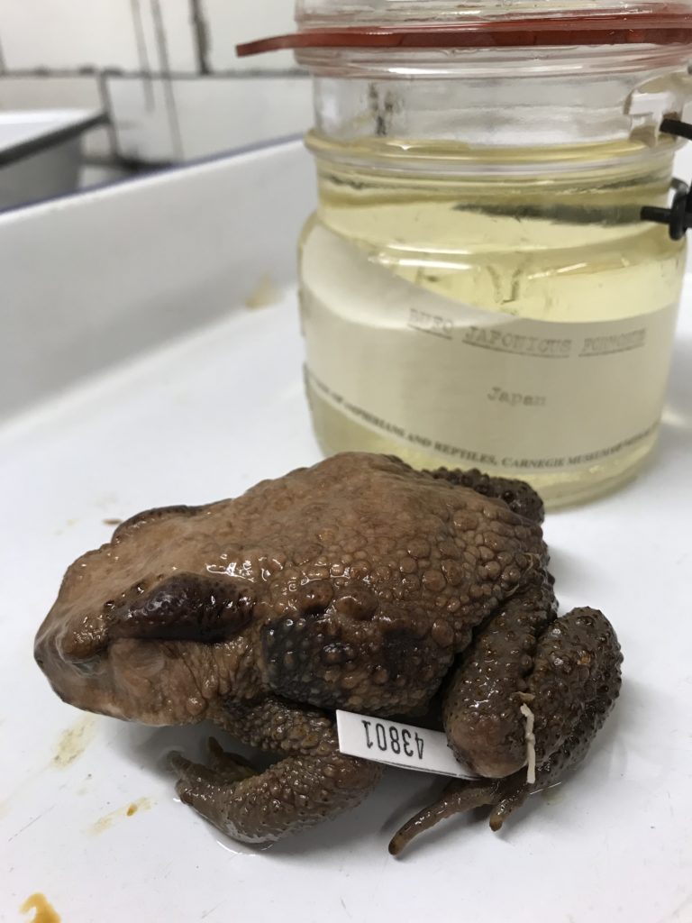
preserved frog specimen
Figure 1. Bufo japonicus. The large glands behind the eyes are called parotid glands, and are a source of toxins in toads. Additionally, all of the bumps you see all over the body are glands that produce skin toxins. Image credit: Stevie Kennedy-Gold.

I recently participated in a Zoom event for Museum members focused on toxins and venom in the natural world. Mason Heberling, Assistant Curator of Botany, and Ainsley Seago, Associate Curator of Invertebrate Zoology, spoke about how the organisms they study produce toxins, and how these chemicals impact their environments and other organisms around them. As an ecologist focused on impacts of climate and land use change, I don’t consider myself an expert on toxins and venoms in amphibians and reptiles, but researching my portion of the joint presentation reminded me once again why herps are the best.

First, a refresher: toxins are poisons, and they have to be consumed or encountered (touched) by an organism to do harm. For example, many frogs produce toxins in their skin, but you would have to either consume that frog or touch its skin for the toxin to do you any harm. Most frogs don’t produce toxins strong enough to hurt humans, though a few notable exceptions exist. Some species of poison dart frogs have skin toxins strong enough that if you touched them and then touched your eyes, nose, or mouth, or if you had a cut on your hand, you could indeed become very ill and perhaps die. Venom, on the other hand, is a toxin that one organism can inject into another. Typically, we think of snakes when we think of injectable toxins. Many snake species have venom glands that produce toxins, and they can forcibly inject that toxin into their prey. The action, which can occur in a flash, involves the use of fangs to puncture the skin, and muscles surrounding the venom gland to force the toxin out along the fang and into the other organism.

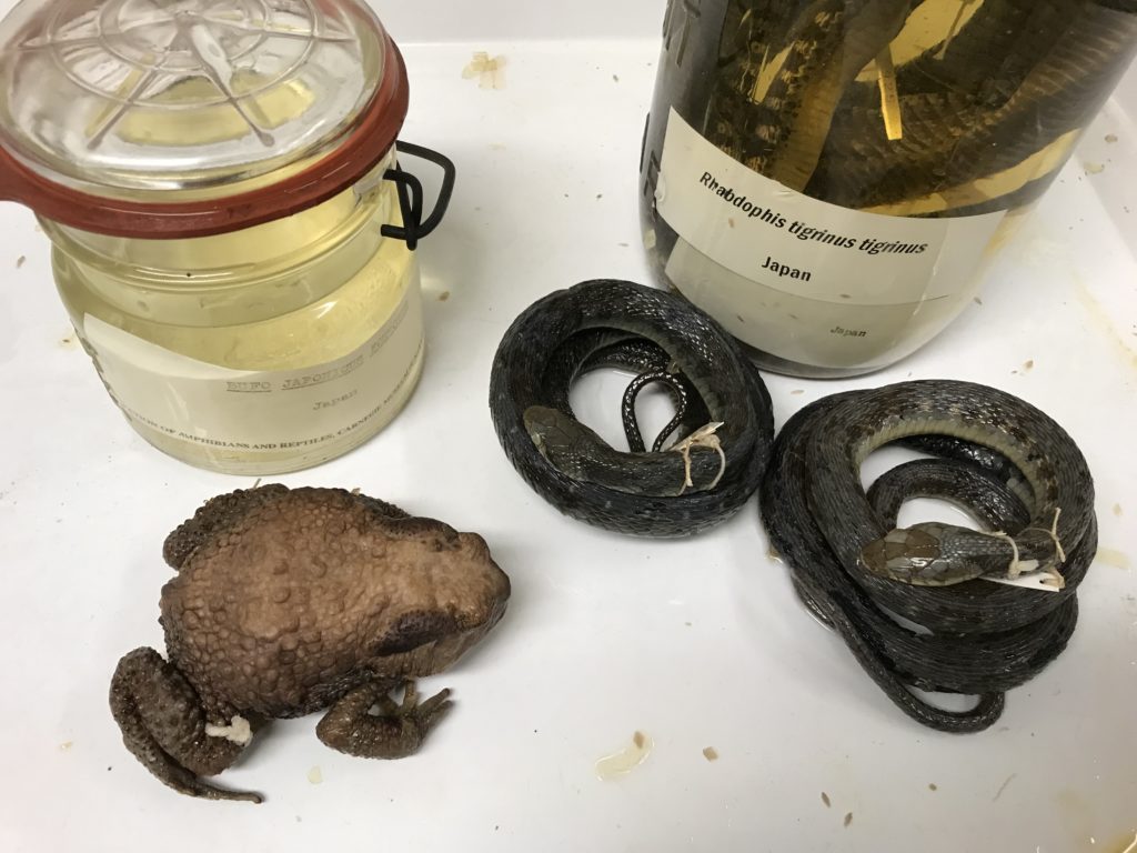
preserved frog specimen, two preserved snake specimens, and two specimen jars
Figure 2. Bufo japonicus and Rhabdophis tigrinus. Rhabdophis are one of the only snakes that are both venomous and toxic. They sequester toxins from the toads they eat into a gland called the nuchal crest. Image credit: Stevie Kennedy-Gold.

Most people tend to think of amphibians as toxic and snakes as venomous. This is true, but it turns out that snakes aren’t the only venomous reptiles, and amazingly two frogs are known to be venomous. Gila monsters (Helodermatidae) and water monitors (Varanidae) produce venom, but their venom glands are in their lower jaws (unlike snakes whose venom glands are in their upper jaws), and they lack the muscles to forcibly inject that toxin the way snakes do. Instead, the act of chewing on their prey causes their jaw motion to work the venom toward their grooved teeth, which then enables the venom to be injected through the bite wound. Using a very different delivery system, two frogs in the family Hylidae (tree frogs from the Americas) have very spiny skulls. Their skin produces toxins, and by “head-butting” another organism, they can effectively inject that toxin into another organism. This unusual delivery system technically makes them both toxic (the toxin can be transferred to you if you touch their skin) and venomous (they can inject that toxin into you).

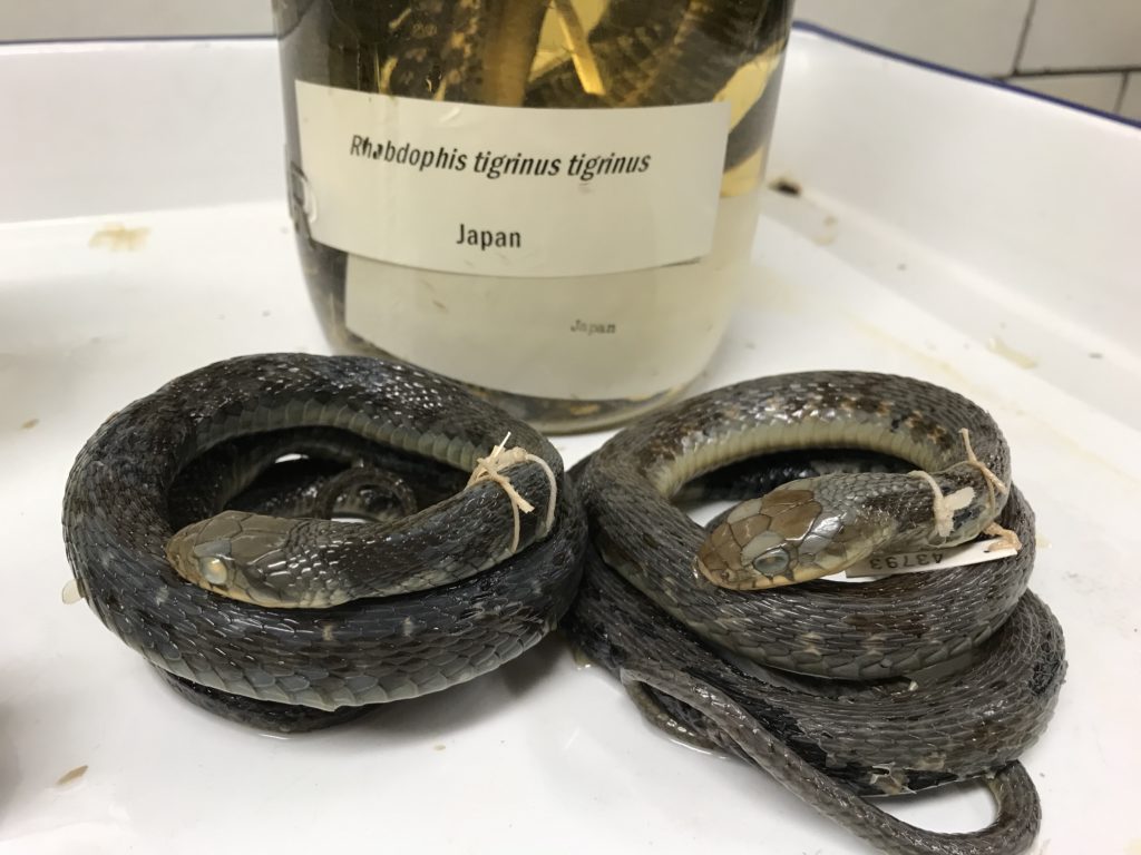
two preserved snake specimens and one specimen jar
Figure 3. Rhabdophis tigrinus. If you look closely at the back of the neck just behind the head on the snake on the left, you can see a slightly raised bit of skin, which is the nuchal crest used to store toxins sequestered from toads. Image credit: Stevie Kennedy-Gold.

One of the most surprising things I learned is that there are snakes that are both toxic and venomous, and these are snakes I see frequently in the field. The genus Rhabdophis is common across South and Southeast Asia, and have long been known to be venomous. What I didn’t know is that in addition to making their own venom, they sequester toxins from their prey, and store it in a gland on the back of their neck called a nuchal crest. Rhabdophis feed on toads, which are toxic, and the snakes are able to sequester that toxin, rather than being adversely affected by it. Interestingly, scientists have shown that Rhabdophis tigrinus are toxic only where their range overlaps with Bufo japonicus, a highly toxic toad—so on some islands of Japan the snakes are toxic, while on other islands they are not.

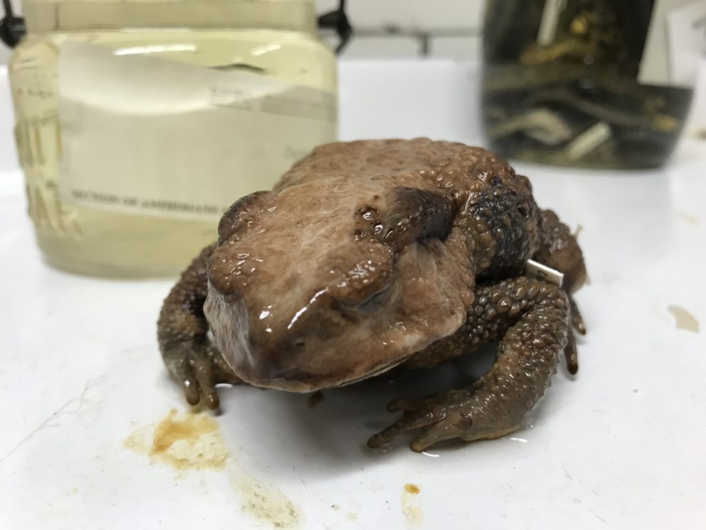
preserved toad specimen
Figure 4. Bufo japonicus with its many toxin glands! Image credit: Stevie Kennedy-Gold.

There are numerous other interesting adaptations involving toxin and venom in the herp world—tweet me (@JenASheridan) if you want to learn more!

Jennifer Sheridan is Assistant Curator in the Section of Amphibians and Reptiles at Carnegie Museum of Natural History. Museum employees are encouraged to blog about their unique experiences and knowledge gained from working at the museum.
Related Content

Early Bats: Ancient Origins of a Halloween Icon

Invasion of the Leaf Galls! 

Jumping Spiders with Sebastian 

Filed Under: Blog Tagged With: booseum, halloween, Jennifer Sheridan, Science News, Super Science Days

October 13, 2020 by wpengine

Early Bats: Ancient Origins of a Halloween Icon

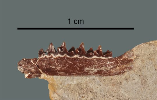
Specimen Carnegie Museum (CM) 62641, the holotypic, or name-bearing, right dentary (lower jaw bone) of the tiny fossil bat Honrovits tsuwape in lingual (= internal) view, still partially encased in ~50-million-year-old rock of the Wind River Formation of west-central Wyoming. Note the length of the scale bar, only 1 cm (less than half an inch)!

Did you know that bats have been around for at least 55 million years? In 1992, several fossils in the Carnegie Museum of Natural History collection, including the lower jaw bone shown above, were described as representing a new genus and species of ancient bat, Honrovits tsuwape—Shoshone for “bat” and “ghost,” respectively—by a team that included two former curators in the museum’s Section of Vertebrate Paleontology, Christopher Beard and Leonard Krishtalka, both now of the University of Kansas. Honrovits dates to the early part of the Eocene Epoch of the Cenozoic Era (the ‘Age of Mammals’), about 50 million years ago, and is a member of a now-extinct bat group called the Onychonycteridae.

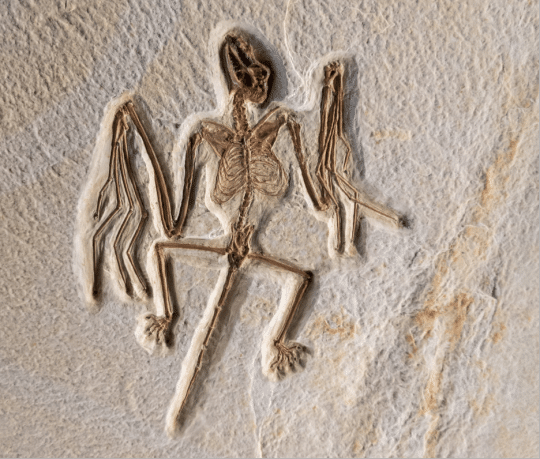
Replica of a beautifully preserved fossil skeleton of Onychonycteris finneyi, a close relative of Carnegie Museum of Natural History’s own Eocene-aged bat Honrovits tsuwape, on display at Fossil Butte National Monument in Wyoming. Photo by Matthew Dillon.

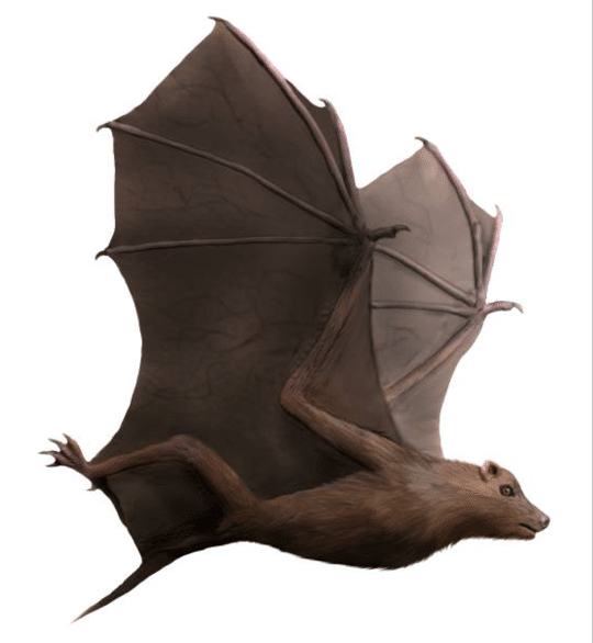
Interestingly, Honrovits shares dental characteristics with a mammal group known as insectivores, which includes today’s hedgehogs, shrews, and moles, and in that sense, it differs from the condition in most other bats. However, bat teeth possess distinctive diagnostic features, so although Honrovits is known only from a few tooth-bearing jaw bones and a skull fragment, there’s no doubt that the diminutive beast was indeed an early bat. The fragmentary nature of its fossils means that we don’t know for sure what Honrovits looked like in life, though it’s a good bet that it bore a close resemblance to other onychonycterid bats, such as Onychonycteris finneyi, which is known from exquisitely preserved skeletons (such as the one shown above).

Flesh reconstruction of the ~50-million-year-old bat Onychonycteris finneyi. There’s an excellent chance that Honrovits tsuwape would have looked like this. Art by Nobu Tamura.

The incompleteness of the Honrovits fossils is, unfortunately, the norm rather than the exception when it comes to prehistoric bats. Fossils of these creatures are exceedingly rare because most bats have very small, light skeletons and achieve their greatest diversity and abundance in areas that have low potential for fossil preservation, such as tropical forests. Occasionally, complete skeletons such as those of Onychonycteris are found, but not nearly as often as fragments.

So, this autumn, if you happen to catch a glimpse of a bat silhouetted against the evening sky, acrobatically wheeling and plunging in pursuit of flying insects, pause and reflect on the history of these extraordinary flying mammals whose ancestry dates nearly to the time of the dinosaurs.

Linsly Church is a Curatorial Assistant in the Section of Vertebrate Paleontology at Carnegie Museum of Natural History. Museum staff, volunteers, and interns are encouraged to blog about their unique experiences and knowledge gained from working at the museum.
Related Content

Halloween and Birds

How Do You Preserve a Giant Pumpkin?

Ask a Scientist: What is the newly discovered “Crazy Beast?”

Filed Under: Blog Tagged With: halloween, Linsly Church, mammals, Science News, Vertebrate Paleontology

October 8, 2020 by wpengine

How Do You Preserve a Giant Pumpkin?

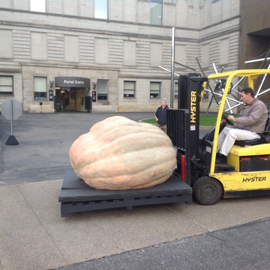
giant pumpkin being moved with a forklift

A few years ago, I came across a dilemma that I wasn’t sure how to resolve. The Section of Botany was given permission to preserve, for the scientific collection, part of the giant pumpkin that was in the exhibition, We Are Nature: Living in the Anthropocene. This was an intriguing offer. I just wasn’t sure how to go about it. Preserving any large fruits or plant parts can be a real challenge. Plant materials must be dried before they rot, and the process must happen at a temperature low enough to prevent the material from being cooked. The normal procedure of putting a plant or plant part into a plant press and drying it with warm dry air was not really an option; at least not for a 2,090-pound pumpkin that wouldn’t even fit in my car, let alone my plant press.

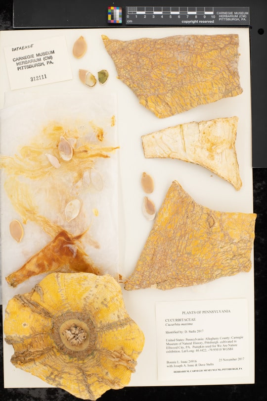
Pumpkins are a type of squash, but trying to literally squash one to dry it seemed a bit daunting. The farmers who grew this giant pumpkin were more than willing to give us whatever parts of the pumpkin we wanted to preserve, and they were even willing to help with cutting them from the pumpkin. We decided on trying to keep the unique parts of the pumpkin, like the stem and the blossom end (bottom). We also saved some of the inner tissue and a few seeds. The seeds on a pumpkin this large are a prize commodity. If a pumpkin from which seeds are properly harvested was a champion, as this one was, each seed could sell for $30 to upwards of $50. It was very generous of the farmers to allow us to have some of these seeds for our collection.

dried pieces of a pumpkin on an herbarium sheet

Pumpkin farmers keep close tabs on the genetics of these giants and actively work at growing larger pumpkins. You can actually find family tree information for this very pumpkin online if you search for it. Who knows how large mankind will eventually enable pumpkins to grow? The plants that grow these large squashes (the Cucurbita maxima variety known as ‘Atlantic Giant’) are a variety of the same species that produce Hubbard Squash. This species, which was originally from South America, has become one of the more diverse domesticated plants.

Giant pumpkins have been a focal point of imagination and literature for some time. Think of Cinderella. There are several variants on the Cinderella tale going back hundreds of years that involve large squash. Back when these stories were written though, it was a fantasy to think there actually could be a pumpkin that a person could fit inside.

Now that we are using QR codes on our herbarium labels, it’s easy to add photographs to plant specimen records. I wish we had thought to do this  before the massive pumpkin was cut up. Maybe I will go back and add a QR code to the label, so the actual pumpkin can be seen again in its full glory. What we have in the collection now are bits and pieces, mere remnants of the gentle giant that grew 45-50 pounds per day in 2017.

Getting back to my original question, how do you preserve a giant pumpkin? I guess the answer is a little bit at a time!

More on this giant pumpkin:

Sasquatch Squash

Giant Pumpkin Seed Harvest 

Collected on This Day: November 25, 2017

Bonnie Isaac is the Collection Manager in the Section of Botany. Museum staff, volunteers, and interns are encouraged to blog about their unique experiences and knowledge gained from working at the museum.
Filed Under: Blog Tagged With: Anthropocene Studies, Bonnie Isaac, Botany, Hall of Botany, halloween, Science News, Section of Botany

October 7, 2020 by wpengine

Halloween and Birds

Birds, being the happy creatures they are, don’t seem to me to connect with Halloween. Sure, death scenes in old movies, or exaggerated depictions of nighttime itself, are often populated with vultures, owls and corvids (crows and ravens), but Halloween itself, not so much. About the only “scary” term I can think of relating to birds is the group popularly referred to as “GOATSUCKERS.”

Early stories about goatsuckers can be credited to Aristotle and Pliny over 2000 years ago. Rumors about a group of birds now classified Caprimulgids, indicated they would suck the milk out of goats, and afterwards the goats would go blind. Of course, the stories are false, but the persistence the common group name might very well continue to frighten young children.

The 70 species of Caprimulgids remain saddled with a Family name, and in some cases a Genus name, that translates from Latin, “capra” for nanny goat, and “mulgēre” to milk, as “milker of goats,” or considering how a bird might attempt such a feat, “goatsucker.”

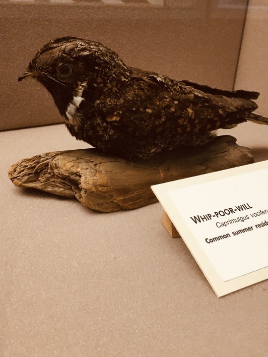
taxidermy mount of whip-poor-will
Image credit: Pat McShea

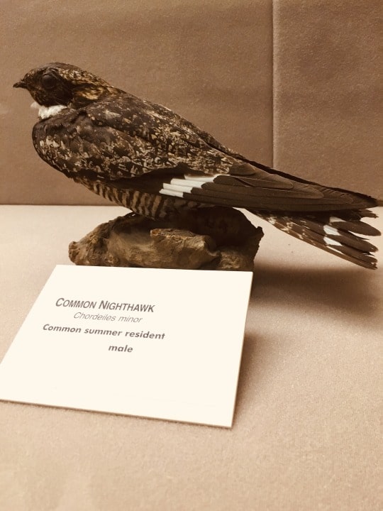
The family Caprimulgidae is a nocturnal group of birds referred to as nightjars or nighthawks that live worldwide except in New Zealand and on some islands in Oceania. In Pennsylvania the only birds of this group seen routinely are the Common Nighthawk and the Whip-poor-will, and both species are declining in numbers. Both are insectivorous birds with what appears to be small mouths that can actually open extremely wide to swallow insects in flight. The sounds of Whip-poor-wills can be haunting to those unfamiliar with them. For an image of the bird and a recording of their distinctive sound click this YouTube link.

taxidermy mount of common nighthawk
Image credit: Pat McShea

The CMNH Section of Birds collection, with nearly 207,000 records, includes only three “goatsuckers” collected on Halloween. Two are Pauraques (Nyctidromus albicollis yucatanensis) from Veracruz, Mexico collected in 1963, and a single Common Nighthawk (Chordeiles minor minor) found dead by former Amphibian and Reptiles Curator Jack McCoy in Schenley Park on Halloween night 1989. Migration should have happened long before that date – in fact this fall Pittsburgh’s estimated peak occurred September 14, when an estimated flight of 50,000 birds of various species passed overhead overnight.

Stephen Rogers is Collection Manager in the Section of Birds at Carnegie Museum of Natural History. Museum employees are encouraged to blog about their unique experiences and knowledge gained from working at the museum.
Related Content

Sympathy For the Devil

Collected on This Day in 1967: Fall Blooms Rival Those of Spring

Ask a Scientist: How do you tell the difference between a raven and a crow?

Filed Under: Blog Tagged With: Birds, Hall of Birds, halloween, Science News, Section of Birds, Stephen Rogers

October 6, 2020 by wpengine

Sympathy for the Devil

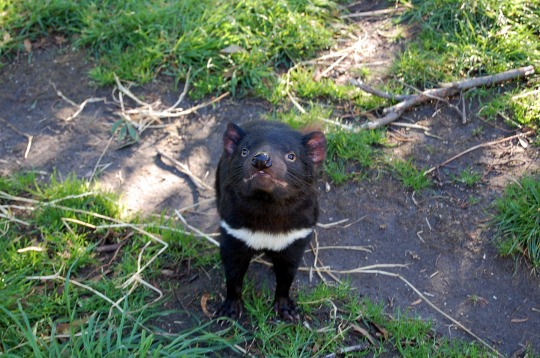
Bats and devils are among the most popular topics associated with Hallowe’en.  Of course, the research collection in the Section of Mammals has worldwide examples of bats species, but we don’t find them scary and we think about bats and their vital ecological roles all year long.  Perhaps more mysterious and less well-known are the two Devil specimens stored among the wombats, kangaroos, and koalas in our collection.  Even school children have heard about *our* kind of devils.  Yes, the Tasmanian devil (Sarcophilus harrisii) is a marsupial – a pouched mammal, like our opossum – that is found only on the island of Tasmania, located some 140 miles off the southeast coast of Australia.  Fossil evidence tells us that it once lived on the Australian mainland, but it may have been wiped out on the continent by the introduction of the Dingo, Australia’s legendary wild dog.

photograph of Tasmanian devil

The Tasmanian devil is a stocky mammal with short legs, short black fur and a distinctive white throat patch. Its head is noticeably large for the size of the body. An adult male may weigh up to 20 lbs. They are nocturnal with a good sense of sight, smell, and touch. Devils are known to cover significant distances nightly, in search of carrion or prey. They can move surprisingly fast and seem to enjoy swimming. In the wild, individuals can live between five and seven years, but many die within the first year of birth. Although it is the largest living marsupial carnivore, the Tasmanian devil is predominantly a scavenger.

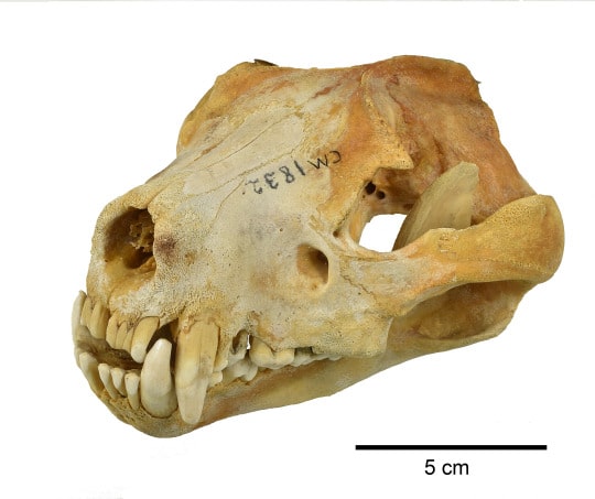
Tasmanian devil skull

A close look at the skull shows evidence of space on the side of the head for large jaw muscles. For its size, the Tasmanian devil has the strongest bite force of any mammal – more powerful than even a hyena! With the large masseter muscles and especially large molars, it can easily crush bone. In fact, devils are such efficient carrion-eaters that they willingly consume an entire carcass, including the fur.

Although this animal gained a reputation for having a bad disposition, it is speculated that this impression was derived from the poor conditions it was kept in when first captured for observations. Since then, it sometimes has been kept humanely as a pet and been found to be much friendlier than initially reported. Tasmanian devils do not seek each other’s company except during the mating period. However, they often come together to feed on a dead animal, where vocalizations and as many as nineteen different behavioral cues are used for communication. These communal gatherings are characterized by aggression and loud sounds, described as “frequent growling” and “blood-curdling screams”!

In 1996, a sad chapter began in the existence of the Tasmanian devil. A deadly infectious cancer called devil facial tumor disease, began to spread within the population. In 2012, the Australian government transferred 30 disease-free individuals to tiny Maria Island off the coast of Tasmania, in what was called ‘island insurance’, while researchers worked on perfecting a vaccine. By 2017, the disease had led to a 90% extinction rate on Tasmania. In hopeful news, by 2019 there were indications that surviving individuals’ immune systems may be undergoing modifications to fight the disease. In early September 2020, a consortium of conservation groups released 11 Tasmanian devils to a wildlife sanctuary in the state of New South Wales, placing the Tasmanian devil on the Australian mainland for the first time in more than 3000 years.  An additional 15 devils were released in early October and more releases are planned.

Currently, the Tasmanian devil is not extinct, but its recovery hangs in the balance. It would be tragic if we are left only with museum specimens and Taz, the Looney Tunes cartoon image, of this fascinating mammal.

Suzanne B. McLaren is the Collection Manager in the Section of Mammals at Carnegie Museum of Natural History. Museum employees are encouraged to share their unique experiences from working at the museum.
Related Content

Scientists Live: Mammalogist Suzanne McLaren on Bats

Vampire Squid: Cutest Dracula

Do Snakes Believe in the Tooth Fairy?

Filed Under: Blog Tagged With: mammals, Museum from Home, Science News, Section of Mammals, Suzanne McLaren

  • « Go to Previous Page
  • Page 1
  • Interim pages omitted …
  • Page 26
  • Page 27
  • Page 28
  • Page 29
  • Page 30
  • Interim pages omitted …
  • Page 43
  • Go to Next Page »

sidebar

About

  • Mission & Commitments
  • Directors Team
  • Museum History

Get Involved

  • Volunteer
  • Membership
  • Carnegie Discoverers
  • Donate
  • Employment
  • Events

Bring a Group

  • Groups of 10 or More
  • Birthday Parties at the Museum
  • Field Trips

Powdermill

  • Powdermill Nature Reserve
  • Powdermill Field Trips
  • Powdermill Staff
  • Research at Powdermill

More Information

  • Image Permission Requests
  • Science Stories
  • Accessibility
  • Shopping Cart
  • Contact
  • Visitor Policies
Stay in the loop! Sign up for our newsletter(s).
One of the Four Carnegie Museums | © Carnegie Institute | Privacy Policy | Terms of Use | Accessibility
Rad works here logo