• Skip to primary navigation
  • Skip to main content
  • Skip to primary sidebar

Carnegie Museum of Natural History

One of the Four Carnegie Museums of Pittsburgh

  • Visit
    • Buy Tickets
    • Visitor Information
    • Exhibitions
    • Events
    • Dining at the Museum
    • Celebrate at the Museum
    • Powdermill Nature Reserve
    • Event Venue Rental
    • Gift Cards
  • Learn
    • Field Trips
    • Educator Information
    • Programs at the Museum
    • Bring the Museum to You
    • Guided Programs FAQ
    • Programs Online
    • Climate and Rural Systems Partnership
  • Research
    • Scientific Sections
    • Science Stories
    • Science Videos
    • Senior Science & Research Staff
    • Museum Library
    • Science Seminars
    • Scientific Publications
    • Specimen and Artifact Identification
  • About
    • Mission & Commitments
    • Directors Team
    • Museum History
  • Tickets
  • Give
  • Shop

Blogs from our Scientific Researchers

Carnegie Museum of Natural History is home to active research and vast scientific collections. Our scientific researchers regularly contribute to the blog at the museum.

October 2, 2020 by wpengine

Vampire Squid: Cutest Dracula

The Vampire Squid is your go-to mollusk for Halloween. It’s covered with glow-in-the-dark spots, and it can hoist its cape-like webbed arms over its head to transform into a pumpkin shape complete with outward-pointing fleshy spines. But wait, there’s more. With the largest eyes relative to body size of any animal, this has got to be the cutest Dracula you ever saw. And the scientific name, inspired by the cloak-like webbing and the dark body color, literally translates to “vampire squid from hell.”
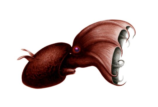
Vampire Squid, showing cloak-like webbing between arms, large eye, and ear-like fins. [Image from Wikimedia Commons]

The Vampire Squid (Vampyroteuthis infernalis) is an extreme deep-water cephalopod more closely related to octopuses than to squids. It is so bizarre that scientists classify it in its own taxonomic order, Vampyromorphida, to show that it differs markedly from other living cephalopods. Like octopuses, it has 8 arms with webbing between them, but unlike octopuses that have suckers on the entire length of the arms, the Vampire Squid bears suckers only on their outermost half. The prominent feature on the arms of the Vampire Squid are fleshy spines or cirri. In addition to the eight arms, it has two velar filaments, in pouches in the webbing, that are analogous (and maybe homologous) to the two long tentacles of squids.

Regarding superlatives, the Vampire Squid has the largest eyes relative to its body size of any other animal, a detail noted in the Guinness World Records. A fully-grown individual can be 28 cm (11 inches) long with eyes 2.5 cm (1 inch) in diameter. Adding to the cuteness factor, they have adorable ear-like fins, which adults use for swimming; juveniles also have fins, but primarily use jet propulsion to move around.

They live in the lightless ocean depths 600-900 m (2000-3000 feet) deep in temperate and tropical oceans world-wide. The ocean at these depths is an oxygen minimum zone with so little dissolved oxygen that most complex organisms cannot survive. But the vampire squid survives perfectly well with a low metabolism and blue blood that is more efficient at carrying oxygen than that of other cephalopods. They use ammonium in their tissues to regulate their buoyancy (ammonium is a wee bit lighter than water), reducing the need for active swimming. Living in the oxygen minimum zone probably helps it to avoid predators.

If disturbed, the Vampire Squid kind of turns itself inside-out into the “pumpkin” or “pineapple” posture by curling its arms and webbing up to cover the body with the spiny cirri pointing outward. Their body is covered by photophores, or light-emitting organs, which they can use to flash a wide range of patterns. In the pumpkin pose, they conceal most of the photophores, but they can light up the tips of the arms and wave them around to distract predators. If it gets really annoyed, the Vampire Squid can release a sticky cloud of luminous mucus that glows for nearly 10 minutes, presumably long enough for the Vampire Squid to make a get-away into the inky darkness.
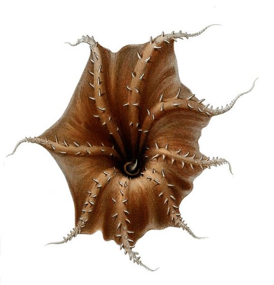
Vampire Squid, underside of arms showing fleshy spines. [Image from Wikimedia Commons]

Much of what we know about their behavior comes from videos made by Remotely Operated Vehicles. It is hard to keep Vampire Squids alive in aquariums at the much lower pressure of our human world, but the Monterey Bay Aquarium succeeded for a while and has some great videos. Aquarium scientists were able to solve the mystery about what the Vampire Squid eats. No, it doesn’t eat blood! It eats detritus (organic debris), also known as marine snow. As the Vampire Squid drifts in the current, any debris that touches an extended filament is moved by the creature’s arms to its mouth. Unusual for being the only known cephalopod to eat non-living food, the Vampire Squid is adapted to eat material that falls through the oxygen minimum zone. Marine snow includes dead bodies, feces, and a lot of mucus from above, and because of the mucus, it is sometimes jokingly referred to as marine snot.

I imagine if Dracula learned about the Vampire Squid, he might exclaim, “I thought it was eating blood, but it’s snot!”

Timothy A. Pearce, PhD, is the head of the mollusks section at Carnegie Museum of Natural History. Museum employees are encouraged to blog about their unique experiences and knowledge gained from working at the museum.

Related Content

Extremely Rapid Evolution of Cone Snail Toxins
Ask a Scientist: Are slugs and snails the same thing?

Chasing Snails in the Great Smoky Mountains

Filed Under: Blog Tagged With: Museum from Home, Science News, Section of Mollusks, Tim Pearce

October 2, 2020 by wpengine

Do Snakes Believe in the Tooth Fairy?

When a child loses a baby tooth, the Tooth Fairy will sneakily appear a short time later to snatch that tooth up and leave behind a little treat. But what happens when vipers or other snakes with large fangs lose their teeth? I doubt the Tooth Fairy would be too keen about sneaking up on a sleeping snake…and as someone who studies and admires snakes, I would not recommend it!

image
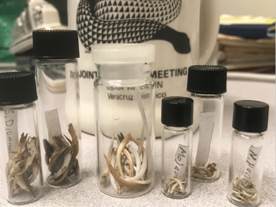
Vials containing Viperidae snake fangs. The middle vial (with clear lid) contains fangs of the Gaboon viper.

I recently learned the answer to this question when I was given seven tiny vials containing dozens of fangs. These fangs came from various species of snakes in the Viperidae family, including the Mojave green rattlesnake (Crotalus scutulatus), the Northern Pacific rattlesnake (Crotalus oreganus), and the Gaboon viper (Bitis gabonica), which has the longest fangs of any snake species. The researcher who gifted us these fangs was curious about the outcome of shed snake teeth and wanted to determine how frequently snake fangs may be swallowed and passed through the snake’s digestive tract. To answer this question, he dissected and examined dried snake feces for the possible presence of shed fangs.*

As it turns out, snakes will occasionally swallow their shed fangs! Vipers are carnivores that have to hunt down and subdue live prey in order to eat and survive. Often there is a struggle between predator and prey and, in that process, a fang may be wiggled lose. Instead of falling out of a snake’s mouth, the snake may swallow the fang along with the prey item. The fang will ultimately pass through the snake’s digestive system and emerge in its feces.

image
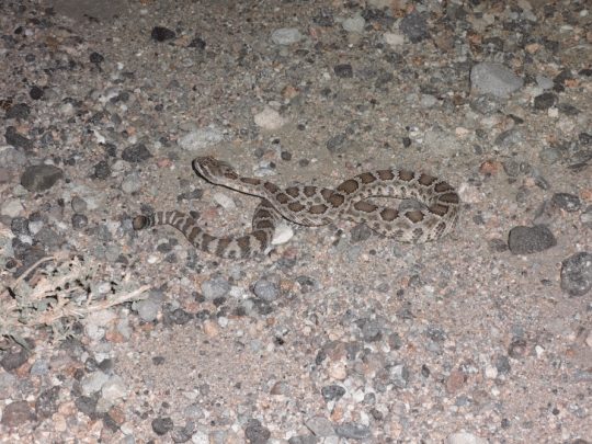
Pacific rattlesnake (Crotalus oreganus) in the Mojave Desert, California. One of the vials contained fangs from this species, but from snakes in the northern portion of the species’ range.

As the collection manager of the Section of Amphibians and Reptiles, I oversee and care for the museum’s massively beautiful and useful collection of cool and creepy herpetofaunal specimens. These specimens include full body wet specimens preserved in alcohol, the osteology collection of bones and turtle shells, and other items such as histology slides, gut contents, and even fangs. Regardless of their preservation form, all the reptile and amphibian specimens within the collection are useful for researchers and could serve to answer future scientific questions. Although the fangs within these seven seemingly unimposing vials have already answered one burning scientific question, they will be added to the collection so see what other answers they can provide!

*Researchers take careful precautions when handling feces as it can carry disease. Do not handle feces you may see in the wild.

Stevie Kennedy-Gold is the collection manager for the Section of Amphibians and Reptiles at Carnegie Museum of Natural History. Museum employees are encouraged to blog about their unique experiences and knowledge gained from working at the museum.

Related Content

Ask a Scientist: What is an Alcohol House?

The Enduring Appeal of Snakes

Poisons of the Carnegie: Timber Rattlesnake

Filed Under: Blog Tagged With: herpetology, Museum from Home, Science News, Stevie Kennedy-Gold

October 1, 2020 by wpengine

The Bromacker Fossil Project Part X: Tambaroter carrolli, an amphibian with a wedge-shaped head

New to this series? Read The Bromacker Fossil Project Part I, Part II, Part III, Part IV, Part V, Part VI, Part VII, Part VIII, and Part IX. 

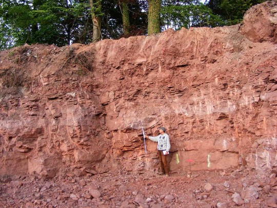
Thomas Martens at the construction site for a new store in Tambach-Dietharz where he found fossils by checking loose pieces of rock on the excavation floor. Photo by Stephanie Martens, 2008.

Paleontologist Thomas Martens has an amazing ability to find fossils. After he discovered the first vertebrate fossils at the Bromacker site in an abandoned commercial quarry in 1974, he and his father Max found additional fossils in the bottom of a deep pit they’d dug with hand tools, an excavation that Dave Berman, Stuart Sumida, and I fondly dubbed the “elevator shaft.” Years later, Thomas used funding from the German federal government to drill rock cores in the field surrounding the Bromacker quarry to help understand the geology of the fossil deposit. Amazingly, at one of the spots Thomas had selected, the drill core penetrated a skeleton of Diadectes absitus. So, it wasn’t surprising that in 2008 Thomas found a skull and partial skeleton of D. absitus and a small skull of a fossil animal new to science at a construction site for a new store in the nearby village of Tambach-Dietharz.

Dave Berman (left) and Stuart Sumida (right) pose with a shopping cart in front of the Netto Discount Store, which was built in the excavation site where Tambaroter was found. Rocks of the Tambach Formation can be seen behind the retaining wall. Photo by the author, 2008.

It makes sense, however, that vertebrate fossils were found close to the Bromacker quarry. Fossils from the Bromacker were preserved in the Tambach Formation, a 200–400-foot-thick unit of sediments that were deposited in the small intermontane Tambach Basin about 290—283 million years ago during the Early Permian Epoch. The Tambach Basin covered an area of about 155 square miles and was internally drained; that is, there were no rivers or streams flowing into and out of the basin. During periods of extremely heavy rain, water and mud would flow down the basin sides in what are called sheet floods and pool in the basin center, which is where the present day Bromacker quarry and Tambach-Dietharz are thought to be located. Any animals killed during these events would be carried by the sheet floods to the basin center where they’d have been quickly and deeply buried in mud settling out of the ponded water and later become fossilized. It is assumed that animals captured by the sheet flood events inhabited the Tambach Basin, because carcasses couldn’t have been carried into the basin by rivers and streams.

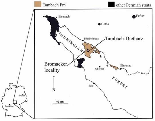
Map of Germany with inset showing the Bromacker locality and the nearby town of Tambach-Dietharz. Although the Tambach Basin in which the Tambach Formation was deposited covers about 155 square miles, outcrops of the Tambach Formation today occur in an area of only about 31 square miles.

While preparing Bromacker fossils, I’d typically read literature related to the fossil I was working on, write notes on what I thought were important features in the fossil, and give my notes to the person leading the project. When Dave was the lead, we’d typically have lots of discussion about certain features preserved in the animal, conversations that often directed the course of preparation. This time, in addition to preparing the new find, I was designated as the lead author for the publication that would name and describe it.

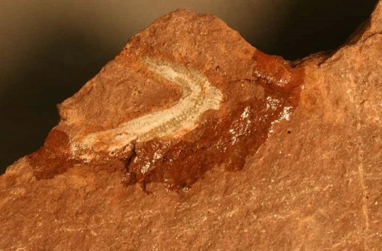
View of the underside of the skull of Tambaroter carrolli before preparation. The shiny area surrounding the skull is glue, which I applied to a crack to stabilize the specimen before preparation could begin. I had to free the skull from the surrounding rock before exposing as much of it as possible through preparation. Photo by the author, 2008.

Tambaroter is a member of the Microsauria, a diverse group of small amphibians that were once thought to be reptiles, a hypothesis that some paleontologists are currently revisiting. Microsaurs inhabited a variety of habitats and exhibited a range of body forms. Some were highly terrestrial with limb proportions similar to those of lizards, whereas others were aquatic and had elongated bodies and reduced girdles and limbs. Still others were adapted for burrowing or rooting through leaf litter. Tambaroter belongs to this latter-most group, which is named Recumbirostra for their recurved snout, in which the front of the mouth is overhung by the snout.

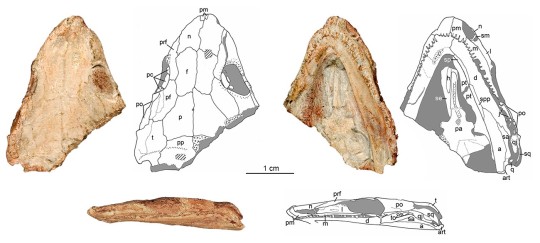
Photographs and line drawings of the skull of Tambaroter carrolli in (clockwise from upper left) dorsal (top), ventral (underside), and left lateral (side) views. Photographs by the author, 2008 and drawings by the author and modified from Henrici et al., 2011.

Tambaroter is member of the recumbirostran subgroup Ostodolepidae. I coined the name Tambaroter, which is derived from “Tamb,” for the Tambach Formation, and the Greek “aroter,” meaning plowman, in reference to the snout shape. Two previously named ostodolepids, Micraroter and Nannaroter, have the “aroter, suffix in their name, so usage of the “aroter” suffix was a continuation of this. The species name, carrolli, honors microsaur expert Robert Carroll (then Curator Emeritus at the Redpath Museum, McGill University, Montreal, Canada).

Skulls of representative ostodolepid microsaurs from geologically oldest (left) to youngest (right). A reconstruction drawing of the skull of Tambaroter was used instead of a photograph for comparison because the original fossil skull is extremely flattened (see previous image). Photographs, except for that of Nannaroter, by the author, 2009. The photograph of Nannaroter was modified from Anderson et al., 2009. Tambaroter skull reconstruction by the author and modified from Henrici et al., 2011. Scale bar of the tiny Nannaroter and other ostodolepids equals 1 cm.

When Tambaroter was published on in 2011, it was the first ostodolepid to be found outside of the USA (the others are from Oklahoma and Texas) and is the oldest one known. Other, possible ostodolepids have since been described from the American Midwest and Germany. All ostodolepids have a wedge-shaped skull and recumbent snout, which is accentuated in Pelodosotis. Based on these features, scientists think that ostodolepids burrowed or searched for worms and other prey in leaf litter. Remarkably, the skull of the tiny Nannaroter is so strongly built that it could have withstood burrowing headfirst into the ground by using its shovel-like snout to loosen dirt and its broad, flat head to push soil against the burrow ceiling. Because the sutures between individual skull bones in the Tambaroter type specimen are not tightly fused together, we think it belonged to a juvenile, so we don’t know if the adult skull would’ve been as strongly built as that of Nannaroter.

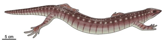
Life drawing of the ostodolepid microsaur Pelodosotis elongatum, which is known by a nearly complete specimen. Tambaroter probably had a similar body shape, though its skull would not have been as strongly wedge-shaped. Drawing modified by Carnegie Museum of Natural History Scientific Illustrator Andrew McAfee from outline drawing in Carroll and Gaskill (1978).

Stay tuned for my next post, which will feature one of the Bromacker’s top carnivores. To learn more about Tambaroter, read the publication that described the animal here.

Amy Henrici is Collection Manager in the Section of Vertebrate Paleontology at Carnegie Museum of Natural History. Museum employees are encouraged to blog about their unique experiences and knowledge gained from working at the museum.

Keep Reading

The Bromacker Fossil Project Part XI: Dimetrodon teutonis, an apex predator

Filed Under: Blog Tagged With: Amy Henrici, Museum from Home, Science News, The Bromacker Fossil Project, Vertebrate Paleontology

September 29, 2020 by wpengine

Collected on this Day in 1967: Fall blooms rival those of spring

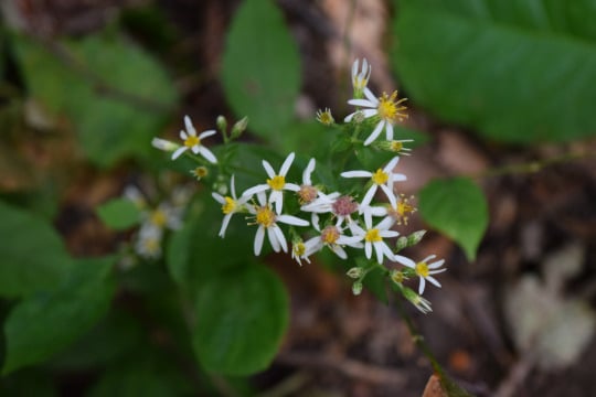
photo of aster flowers with white petals

In the northeastern United States, we often think of spring as a time for wildflowers.  But the fall is, too.  

It is easy to be distracted by the beautiful fall foliage, when our landscape turns brilliant shades of red, orange, and yellow.  But when many plants are shutting down for the winter, others are just kicking into gear.

Many wildflower species bloom well into fall, both in open areas and in the forest understory.  One group of plants are the fall blooming “asters.”  In same plant family as sunflowers and dandelions (Asteraceae), Aster was once a very large plant genus in our native North American flora (somewhere along the lines of >175 species!), but as we learned more about the evolutionary relationships of these plants, they have since been split into multiple genera (plural of genus). In fact, there is only one “true” Aster in Pennsylvania, Tatarian aster (Aster tataricus), which is actually not even native to Pennsylvania!  Regardless of the scientific name, these plants are commonly referred to as asters.  And they put on quite an autumn show in Pennsylvania.

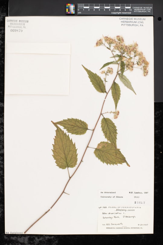
dried specimen of aster flower from Carnegie Museum of Natural History herbarium

Perhaps one of the most common woodland asters in Pennsylvania is white wood aster (Eurybia divaricata, formerly known as Aster divaricatus).  This specimen was collected September 29, 1967 by N.R. Farnsworth in Pittsburgh’s Schenley Park.  This species can still be found in Schenley Park, and many parks, woodlands, and wooded roadsides across Eastern North America.

Fall foliage is beautiful in Pennsylvania.  But don’t forget to look down at the flowers, too!

Find this white wood aster specimen here: https://midatlanticherbaria.org/portal/collections/individual/index.php?occid=11826562

Check back for more! Botanists at the Carnegie Museum of Natural History share digital specimens from the herbarium on dates they were collected. They are in the midst of a three-year project to digitize nearly 190,000 plant specimens collected in the region, making images and other data publicly available online. This effort is part of the Mid-Atlantic Megalopolis Project (mamdigitization.org), a network of thirteen herbaria spanning the densely populated urban corridor from Washington, D.C. to New York City to achieve a greater understanding of our urban areas, including the unique industrial and environmental history of the greater Pittsburgh region. This project is made possible by the National Science Foundation under grant no. 1801022.

Mason Heberling is Assistant Curator of Botany at Carnegie Museum of Natural History. Museum employees are encouraged to blog about their unique experiences and knowledge gained from working at the museum.

Related Content

Collected on This Day in 1944: Squarrose Goldenrod

Dandelion and Goldenrod Coloring Pages

Herbarium Specimens Hold More Information Than We Realize

Filed Under: Blog Tagged With: Botany, collected on this day, Hall of Botany, Mason Heberling, Museum from Home, Science News, Section of Botany

September 28, 2020 by wpengine

Herbarium specimens hold more information than we realize

The first herbarium I visited was the Pringle Herbarium at the University of Vermont as part of an undergraduate class on plant taxonomy and systematics. Prior to this visit, I assumed herbaria were fairly mundane collections of dead, dry, flattened plants, and that they couldn’t possibly interest me as much as emerald-green plants thriving in the wild. However, within moments of entering the Pringle Herbarium, I was captivated by the football-sized cones of the sugar pine (Pinus lambertiana). These giant cones, of a species native to mountain slopes in California and Oregon, were the largest of any gymnosperm I had seen at that time, and I quickly discovered that herbaria were fascinating resources for studying plant diversity around the world.

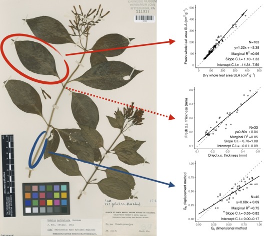
Plant specimens capture important information on plant traits across species, continents, and centuries. With over 390 million specimens worldwide and becoming increasingly available online (500,000 specimens at Carnegie Museum alone), that’s a lot of potential information! We found that measurements using herbarium specimens strongly correlate to those measured in the field, including two leaf traits and one stem trait.

Years later as a graduate student interested in plant functional ecology, I was reminded of the diversity contained within herbaria, but learned that herbarium specimens were rarely used to study plant functional traits. Functional traits are characteristics that provide ecologists with information about growth, reproduction, or survival strategies, and in plants they are often measured using living tissue. For example, three commonly measured functional traits are specific leaf area, wood density, and leaf thickness. Specific leaf area (equal to the fresh area of a leaf divided by its dry mass) indicates how much dry mass plants invest in their leaves, a factor coordinated with their rate of photosynthesis. More specifically, plant photosynthetic rates tend to increase the bigger leaves get relative to their dry mass. On the other hand, wood density is used to understand carbon storage, which is important for studying carbon sequestration and climate change. Leaf thickness can help understand leaf thermoregulation, herbivory, and gas exchange. Currently, it’s unclear if herbarium specimens can provide reasonable estimates of these traits, but if so herbaria can vastly expand our understanding of plant functional diversity.

Recently, I teamed up with scientists Jessica Rodriguez and Dr. Mason Heberling (Assistant Curator of Botany at Carnegie Museum of Natural History) to understand if and to what extent herbarium specimens could be used as proxies for functional traits collected from fresh plant tissues. In our study just published in the American Journal of Botany, we found that herbarium specimens can provide accurate estimates of specific leaf area, branch wood density, and leaf thickness. Although drying plant tissues may lead to some inaccuracies in functional traits that are typically measured using fresh tissues, our study suggests the dead, dry, flat plants I once considered uninteresting could rapidly advance what scientists know about plant functional diversity. Importantly, our research highlights herbaria as rich sources of functional trait data with the potential to accelerate the study of important ecological processes like species responses to climate change.

Timothy M. Perez, Ph.D. is a postdoctoral scholar at the University of British Columbia whose research focuses on plant heat tolerance and the conservation of plants in the tropics.

Related Content

The Circle of Life…and Invasion

A Couple of Creepers: Plant Doppelgängers

Ask a Scientist: How Do You Find Rare Plants?

Filed Under: Blog Tagged With: Botany, Hall of Botany, Science News, Section of Botany

September 25, 2020 by wpengine

From Collector to Director

image
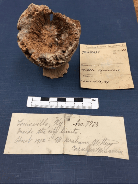
Figure 1: CM 50625 – Rugosa Coral. Collected by M. Graham Netting in 1912.   Coral body shape has a radial symmetry.

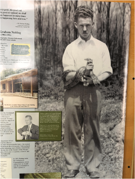
In 1912, eight-year-old M. Graham Netting unearthed 13 coral fossils within the city limits of Louisville, Kentucky.  Later, as a 22-year-old Pitt student, he donated them to the Carnegie Museum of Natural History (Figure 1).   When the Great Depression cut short his graduate studies at the University of Michigan in 1929, he returned to the museum as Assistant Curator of Herpetology, and worked his way up to Curator in 1932.   In 1954, six months before turning 50, he was appointed Director of the Carnegie Museum of Natural History.   Along the way, the Wilkinsburg native left an astonishing legacy that includes a steady growth in scientific collections, numerous wildlife dioramas in the Halls of Wildlife, and a mid-Appalachian field research station, Powdermill Nature Reserve.  Upon his retirement in 1975, the Post-Gazette noted, “Long before it was “in,” Netting saw pollution of the air and water ravaging the land.”

Albert Kollar, Collection Manager of the of Section of Invertebrate Paleontology, re-discovered young Graham Netting’s horn corals while working on a multiyear review of the Bayet Collection.  Netting’s label note did not provide any evidence for the stratigraphic unit that he collected from, but more on that later.

image
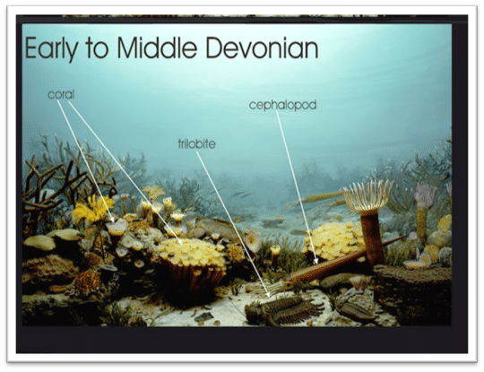
Figure 2: Carnegie Museum of Natural History exhibit reconstruction of an Early to Middle Devonian reef, 375 – 390 MYA.  The reef shows Rugosa and Tabulate corals, a spiny trilobite about 18 inches in length and several straight cephalopods.   Coral tentacles (shown in white) are illustrated in feeding mode.  Both Rugosa and Tabulate corals went extinct at the end of the Permian Period.

Rugose corals are often called horn corals because many species have a horn shape.  Horn corals attach to the sea floor by way of a sticky tentacle that protrudes from the base or curved end of the animal.  Other invertebrate animals, such as brachiopods, attached in this position are described as sessile.  The coral animal or “polyp” built its skeleton from calcium carbonate, a mineral formed from Bicarbonate and Calcium ions in seawater.  The polyp tentacles or feeding polyp extend out from the top of the basic body for feeding (Figure 2).  When the animal died, its soft tissues would have decayed and left behind the external hard mineral skeleton that fossilized.

Netting’s Louisville coral specimens are fossilized in a different way than similar corals from the nearby Falls of the Ohio middle Devonian fossil beds.  His corals are lighter and fragile to the touch, conditions which gave Albert reason to compare Netting’s fossils to similar invertebrate paleontology corals from strata within the Louisville area.  Sometime during or after burial, these horn coral skeletons were replaced by silica or quartz, a process known as silicification. The mineral silica can saturate a column of seawater when the seabed is overwhelmed with a large population of sponges.  Sponge skeletons are composed of silica and when they die silica is added to a column or more of seawater.  Volcanic eruptions eject silica into the atmosphere that eventually settles into the sea.  Again potentially adding higher amounts of silica.  Whatever the cause, Albert believes Netting’s corals were collected from the fossiliferous Middle Devonian age Jeffersonville Limestone, where the “lower foot of a “conglomerate” of reworked silicified Louisville Limestone” of Upper Silurian age is known to occur with silicified coral fossils (Conkin and Conkin, 1972).

Horn and Tabulate corals thrived in shallow seas forming diversified ecological reefs from about the late Silurian Period to the beginning of the Late Devonian epoch. During the Middle Devonian epoch roughly 400 Ma to 390 Ma years ago, reefs formed in central New York, southern Ontario, central Ohio, central Iowa, western Alberta, Canada, western Australia, and in Eifel, Germany.

image
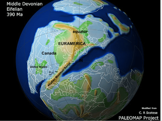
Figure 3:  Paleogeographic Map of the Middle Devonian Period – Kentucky is well south of the equator.

Louisville, during the Devonian Period, was centered in the southern hemisphere about 40 degrees south of the equator. Because of plate tectonics, the coral beds of Louisville would travel 5,500 miles over the next 390 million years to their present-day location of 38 degrees north (Figure 3). Today, fossil outcrops in the city limits of Louisville are difficult to find.

Figure 4: Graham Netting in his twenties.

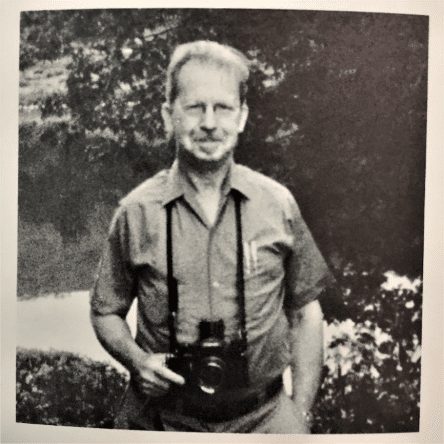
When Netting retired as Director of Carnegie Museum of Natural History, he moved to a modest house next to Powdermill Nature Reserve.   A seat was saved for him each Sunday at the reserve’s weekly nature talk.  In 1996, he passed away.  Steve Rogers, Collections Manager for the Section of Birds, recalls sipping fresh lemonade on Netting’s back porch in 1981.   According to Rogers, Netting was reflective and humble.   The fossil collector who became a museum director had a habit of rubbing his chin while listening to someone speak.   When asked about his legacy, Rogers replied, “He was more instrumental in forming Powdermill than anyone.  He had an amazing ability to be a part of a team that got things done.”

Figure 5: Graham Netting at Retirement in 1975.

As Netting prepared to step down as director in 1975, he said, “These great collections are a natural resource to answer questions about the life of the world.” On a recent day, I saw two children jumping up and down in front of the Glacier Bear diorama in Hall of North American Wildlife on a family visit to the museum.   When one of the children asked, “what’s a diorama?” I thought about Graham Netting, smiled, and encouraged their engagement with the life of the world.

Many thanks to Xianghua Sun, Carnegie Museum Library Manager, Marie Corrado, Carnegie Museum Library Clerk, Stephen Rogers, Collections Manager for the Section of Birds, and John Wenzel, Director of Powdermill Nature Reserve for help researching this post.  

Joann Wilson is an Interpreter in the Education Department at Carnegie Museum of Natural History and Albert Kollar is Collections Manager for the Section of Invertebrate Paleontology. Museum employees are encouraged to blog about their unique experiences and knowledge gained from working at the museum.

Related Content

CMP Travel Program and Section of Invertebrate Paleontology Promote the 125th Anniversary of Carnegie Library of Pittsburgh with an Outdoor Walking Tour

Hidden Treasure of Pittsburgh Found

Harriet Tubman Was a Naturalist

Filed Under: Blog Tagged With: Albert Kollar, Graham Netting, invertebrate paleontology, Joann L. Wilson, Museum from Home, Powdermill Nature Reserve, Science News

  • « Go to Previous Page
  • Page 1
  • Interim pages omitted …
  • Page 27
  • Page 28
  • Page 29
  • Page 30
  • Page 31
  • Interim pages omitted …
  • Page 43
  • Go to Next Page »

sidebar

About

  • Mission & Commitments
  • Directors Team
  • Museum History

Get Involved

  • Volunteer
  • Membership
  • Carnegie Discoverers
  • Donate
  • Employment
  • Events

Bring a Group

  • Groups of 10 or More
  • Birthday Parties at the Museum
  • Field Trips

Powdermill

  • Powdermill Nature Reserve
  • Powdermill Field Trips
  • Powdermill Staff
  • Research at Powdermill

More Information

  • Image Permission Requests
  • Science Stories
  • Accessibility
  • Shopping Cart
  • Contact
  • Visitor Policies
Stay in the loop! Sign up for our newsletter(s).
One of the Four Carnegie Museums | © Carnegie Institute | Privacy Policy | Terms of Use | Accessibility
Rad works here logo