• Skip to primary navigation
  • Skip to main content
  • Skip to primary sidebar

Carnegie Museum of Natural History

One of the Four Carnegie Museums of Pittsburgh

  • Visit
    • Buy Tickets
    • Visitor Information
    • Exhibitions
    • Events
    • Dining at the Museum
    • Celebrate at the Museum
    • Powdermill Nature Reserve
    • Event Venue Rental
    • Gift Cards
  • Learn
    • Field Trips
    • Educator Information
    • Programs at the Museum
    • Bring the Museum to You
    • Guided Programs FAQ
    • Programs Online
    • Climate and Rural Systems Partnership
  • Research
    • Scientific Sections
    • Science Stories
    • Science Videos
    • Senior Science & Research Staff
    • Museum Library
    • Science Seminars
    • Scientific Publications
    • Specimen and Artifact Identification
  • About
    • Mission & Commitments
    • Directors Team
    • Museum History
  • Tickets
  • Give
  • Shop

Enjoy the Museum from Home via our Blog

Can't make it to the museum in person? We've done our best to help cultivate resources for you to enjoy from home. Activities for the whole family, different ways to experience our exhibitions and more are included in these blogs.

August 3, 2020 by wpengine

Cats: The Archeological Site!

There’s an internet meme making the rounds that says if dogs are “man’s best friend” then cats are basically weird roommates. However, if you happened to live in ancient Egypt, you’d consider cats to be tad more special—a veritable link to the divine, in fact. Cats were of great importance in matters both earthbound and spiritual in Egypt, beginning even before the First Dynasty over 5,000 years ago. Aloof but lovable, cats played the role of pet, hunter, and deity in ancient Egypt and to this day they haven’t forgotten. Believe me, cats know.

Let’s start with the practical role that cats played in Egypt. Has yours ever left you a present of a dead mouse or bird? Odds are that it has—whether you liked it or not. Ancient Egyptians valued cats for this very skill. Cats hunted the rodents that threatened to devour Egyptian grain and spread deadly diseases. Cats also hunted animals dangerous to humans like snakes and scorpions. Tomb paintings also depict cats helping their royal owners hunt elusive marsh birds for sport. Egyptians loved cats for their companionship as well—not just as hard-working professionals—and played a major role in domesticating them. Ancient Egyptian art captures cats wearing collars and lurking under chairs not so differently from the cats that keep us company today.

When cats stretched themselves out in the sun for a catnap, ancient Egyptians associated them with the sun god Ra and his daughter Bastet. Bastet was the goddess of the home, fertility, joy, and the protection of children; and she is often depicted in statuary as a woman with the head of an alert, attentive cat. Even earlier depictions of Bastet, however, show a fierce and wild lioness. Some scholars believe this shift in imagery is connected to the domestication of cats—from the Near Eastern wildcat (Felis silvestris lybica) to the modern-day housecat (Felis catus). These traits of the goddess Bastet—vigilance, protectiveness, companionship—were reflected in the characteristics of Felis catus. Ra, in his cat form, also shared these characteristics. When accompanying a deceased Egyptian to the afterlife, Ra was prepared to defend them from Apep, the serpent god of chaos and disorder.
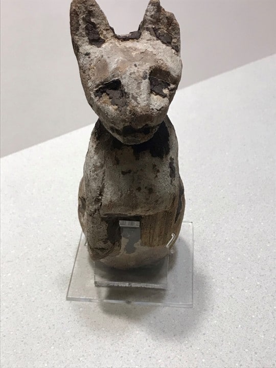
cat statuette

By the Ptolemaic period of Egyptian history (305-30 BCE), Bastet was hugely popular. Her temples drew thousands of pilgrims every year. These pilgrims would buy statuettes of the goddess or actual cat mummies to leave at the temple. This was a way for the pilgrims to commemorate their visit and to venerate Bastet. When the number of these statues and cat mummies grew too large, the priests of the temple would dig special trenches and bury them to make room for more. About two thousand years later in the nineteenth century, archeologists would begin to unearth these trenches and discover more cat mummies and Bastet statuary than they knew what to do with. Unfortunately, some English excavators even sent the cat mummies they discovered back to Britain…to be ground into fertilizer!
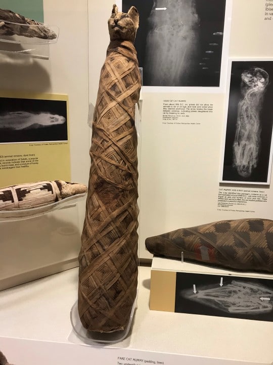
cat mummy in museum display

Millions of cats were mummified in ancient Egypt either to be buried alongside their owner or to be sold to pilgrims devoted to Bastet. Cat mummification in the name of Bastet became an industry because many temples—depending on the whim of pharaonic decree—had to sustain themselves financially on their own. Sometimes a temple might sell a pilgrim a “fake” cat mummy! And it’s one of these curiosities that the Carnegie Museum of Natural History has on display in Walton Hall of Ancient Egypt. While it looks like a standard-issue cat mummy (Roman period, c. 30 BCE), an x-ray led to the discovery that the remains belong to another undetermined animal.
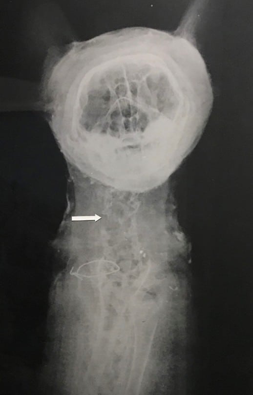
x-ray of cat mummy

Cats played a central role in the daily life and religious practices of ancient Egyptians. They kept their humans safe from snakes and scorpions and Egypt’s grain supply safe from rats and mice. Cats even came to represent in animal form some of Egypt’s most important gods. So, the next time your cat ignores you and wanders off, know that one of its ancestors quite possibly did the same thing to a pharaoh.

Nicholas Sauer is a Gallery Experiences Presenter and Natural History Interpreter at the Carnegie Museum of Natural History. Museum staff, volunteers, and interns are encouraged to blog about their unique experiences and knowledge gained from working at the museum.

Works Cited

“Bastet.” The Louvre Museum. 2009. <https://www.louvre.fr/en/oeuvre-notices/bastet>.

Castellano, Nuria. “The Sacred and Secret Rituals in the Egyptian Book of the Dead.” National Geographic, 8 February 2018. <https://www.nationalgeographic.com/history/magazine/2016/01-02/egypt-book-of-the-dead/>.

Grimm, David. “Ancient Egyptians May Have Given Cats the Personality to Conquer the World.” Science Magazine, 19 June 2017. <https://www.sciencemag.org/news/2017/06/ancient-egyptians-may-have-given-cats-personality-conquer-world>.

Little, Becky. “Kitten Mummies.” History.com, 18 November 2018. <https://www.history.com/news/ancient-egypt-cat-mummy-discovery-scarab>.

Macdonald, James. “Why Ancient Egyptians Loved Cats So Much.” JSTOR Daily, 27 November 2018. <https://daily.jstor.org/why-ancient-egyptians-loved-cats-so-much/>.

“Paintings from the Tomb-chapel of Nebamun.” Khan Academy. 2020. <https://www.khanacademy.org/humanities/ancient-art-civilizations/egypt-art/new-kingdom/a/paintings-from-the-tomb-chapel-of-nebamun>.

Related Content

Meowfest: Museum Scavenger Hunt
Meowfest: Why do Cat Eyes Glow in the Dark?

Stuffed Animal Safari: Jaguars

Filed Under: Blog Tagged With: cats, Museum from Home, Super Science Days, Super Science Meowfest, Walton Hall of Ancient Egypt

July 31, 2020 by wpengine

Mesozoic Monthly: Aspidorhynchus

As we all seek out responsible ways to enjoy our summer months while the world continues to respond to COVID-19, many of us are embracing the therapeutic effects of the great outdoors. One popular activity, especially in and around the Three Rivers, is fishing. Some modern fishes look positively primeval, as if they were hooked straight out of the Age of Dinosaurs and reeled into the present day. For July’s edition of Mesozoic Monthly, our star is Aspidorhynchus, one of the weird and wonderful fishes that inhabited the oceans of the Mesozoic Era.

Let’s start with a quick lesson on fish, for context. There are two main groups of bony fishes. One group, the class Sarcopterygii, are called the lobe-finned fishes because they have fleshy, limb-like fins that they use to paddle through the water like oars. The first vertebrates to go on land were sarcopterygians, and the descendants of these adventurous fish eventually evolved into amphibians, reptiles, and mammals – including us! Despite their prolific limbed descendants, sarcopterygians make up only a small fraction of fishes today. The vast majority of fish belong to the other class: Actinopterygii, or the ray-finned fishes. These fishes have delicate ray-like bones supporting thinly webbed fins instead of the meaty fins of the sarcopterygians. Actinopterygians are so successful that they dominate both freshwater and saltwater ecosystems, thrive in a variety of habitats, and fill various ecological niches. Such diverse lifestyles mean that actinopterygians come in many shapes and sizes. Nemo (a clownfish) is an actinopterygian. So is the barracuda that ate his mother, the catfish in the Monongahela River, and the unfortunate goldfish you won at the carnival as a kid. Most fossil fishes, like Aspidorhynchus for example, are also actinopterygians.

Aspidorhynchus is an extinct member of the order Holostei, nested, in diagrams of relatedness, within the class Actinopterygii. The only members of the Holostei today are gars and bowfins. Superficially, Aspidorhynchus looks like a gar, but it is more closely related to bowfins. Its name means “shield snout,” in reference to its pointy, swordfish-like upper jaw. Unlike swordfish, which lack teeth as adults, this snout was filled with many sharp teeth. The limited flexibility of its skull restricted its diet to tiny fish, two inches (5 centimeters) in diameter at the largest. Aspidorhynchus was not very large itself, its slender body only growing to approximately two feet (60 centimeters) in length. It was covered with ganoid scales, which are hard, diamond-shaped scales made with a shiny compound called ganoin. Only a few types of modern fishes have ganoid scales, including gar, sturgeon, and paddlefish.

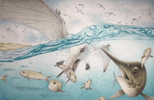
Jurassic feeding frenzy: the pterosaur (flying reptile) Rhamphorhynchus and the predatory fish Aspidorhynchus attack a school of smaller fish. Usually, the baitfish were the only casualties here, but once in a while, everybody lost (see below!). Art by RavePaleoArt on DeviantArt, reproduced with permission.

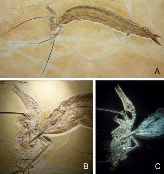
Although species of Aspidorhynchus lived in the Jurassic and Cretaceous periods, we know that it encountered the same struggles as some modern fish due to several remarkable fossils. Just like swordfish, the pointy snout of Aspidorhynchus frequently got it into trouble by impaling other animals! The abundance of fossil evidence for this was provided by the unique conditions of the habitat preserved in the famous Solnhofen Limestone of Germany. In the Late Jurassic, this area was an isolated series of lagoons that accumulated a bottom layer of anoxic brine, which is extra-salty, low-oxygen water where oxygen-dependent (aerobic) life cannot survive. Despite this, the surface still teemed with life: fishes and marine reptiles dominated the water, small non-avian dinosaurs scurried along the shore, and pterosaurs (flying reptiles) and archaic birds flew overhead. The fish-eating pterosaur Rhamphorhynchus seems to have been a fairly frequent victim of the snout of Aspidorhynchus, with multiple fossils documenting unfortunate collisions in which the fish’s snout pierced and became entangled in the wing membrane of the pterosaur. (For a summary of pterosaur wings, check out the March edition of Mesozoic Monthly, on Nemicolopterus.) It’s obvious from the size of the animals that neither was trying to eat the other, but somehow, they became stuck together. As the two animals struggled to survive, they slowly drifted downward into the anoxic brine, where they suffocated and settled onto the bottom of the lagoon. If any other animals had tried to eat or otherwise disturb the corpses, they would have died in the brine as well, so the fossils of the Solnhofen Limestone are typically pristine and undisturbed by scavengers.

Three views of the most famous (and probably the most beautiful) Aspidorhynchus vs. Rhamphorhynchus fossil from the Upper Jurassic Solnhofen Limestone of southern Germany. Avid fisherman Matt Lamanna, the head of Vertebrate Paleontology at Carnegie Museum of Natural History (CMNH), jokes that the Aspidorhynchus looks angry, as if it’s mad about getting its snout stuck in the Rhamphorhynchus and dooming them both. Sorry Matt, this is just a quirk of preservation – the compression of the Aspidorhynchus skull during fossilization gave it the appearance of having grouchy eyebrows that weren’t there in life. You can learn more about this specimen in a paper by Frey and Tischlinger (2012).And if you want to see real fossils of both of these animals in person (albeit preserved separately), come visit the Solnhofen case in CMNH’s Dinosaurs in Their Time exhibition.

Because Aspidorhynchus lived only during the Mesozoic, there’s no chance that a modern-day angler will ever hook one. But should you find yourself fishing in one of Pennsylvania’s rivers or lakes this summer, and manage to land a gar or bowfin, pause for a moment and reflect on the ancient legacy of these fishes – a heritage that dates to the Age of Dinosaurs.

Lindsay Kastroll is a volunteer and paleontology student working in the Section of Vertebrate Paleontology at Carnegie Museum of Natural History. Museum staff, volunteers, and interns are encouraged to blog about their unique experiences and knowledge gained from working at the museum.

Related Content

Preserving Fossil Treasures: Eocene Fishes from Monte Bolca, Italy

Science Chats: Herpetologist Talks About Dinosaurs with Paleontologist

The Bromacker Fossil Project Part VIII: Martensius bromackerensis Honoring a Colleague

Filed Under: Blog Tagged With: dinosaurs in their time, Lindsay Kastroll, Mesozoic Monthly, Museum from Home, Science News, Vertebrate Paleontology

July 30, 2020 by wpengine

The Bromacker Fossil Project Part VIII: Martensius bromackerensis, Honoring a colleague

New to this series? Read The Bromacker Fossil Project Part I, Part II, Part III, Part IV, Part V, Part VI, and Part VII. 

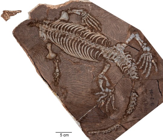
Adult, holotype specimen of Martensius bromackerensis. Image digitally assembled by the author from five photographs taken by Diane Scott (Preparator at University of Toronto Mississauga [UTM]), 2010–2013. The specimen was collected in several large blocks.

The formal publication of some of the Bromacker discoveries took more time to complete than others, and our most recently pubished fossil, Martensius bromackerensis, holds the record in that regard. Four nearly complete specimens of Martensius were collected from the Bromacker quarry between 1995–2006. The first, discovered by Thomas Martens and his father Max, came from a jumbled pocket of fossils. Unfortunately, muddy groundwater had penetrated cracks in the subsurface of this portion of the quarry and coated and eroded bone present along these cracks. Despite this damage and the lack of a skull, we could identify the specimen as a caseid synapsid (synapsids, also known as mammal-like reptiles, are a group of amniotes whose later-occurring members gave rise to mammals).

Drawing of 1995 Martensius bromackerensis specimen. Because the specimen was collected in numerous pieces of rock, with parts of some bones exposed on apposing rocks, Scientific Illustrator Kevin Dupuis (UTM) had to first draw the bones exposed on each piece and then assemble all of the drawings digitally. Dotted lines indicate bone impression in the rock. Arrows point to healing scars from two fractures in the last right rib. Additional healing scars can be seen in preceding ribs. This animal apparently survived a serious injury. Modified from Berman et al., 2020.

The next specimen was discovered in 1999 by Georg Sommers (Preparator, Museum der Natur, Gotha), who prepared the fossil. It consists of a vertebral column, ribs, some limb bones, and a few scattered skull elements. Unfortunately, a more complete skull was needed to allow for comparison to other caseids, some of which are based only on skull material. It wasn’t until the discovery of two more specimens in 2004 and 2006 by Stuart Sumida and Dave Berman, respectively, that the long sought-after skull was found. Preparation of these specimens took a long time due to their size and the considerable amount of rock covering the bones in some of the blocks. My promotion to Collection Manager in 2005 left me with considerably less time to prepare fossils. Other preparators were asked to help with the preparation at both Carnegie Museum of Natural History (CMNH, Dan Pickering and Tyler Schlotterbeck) and in Dr. Robert Reisz’s lab at the University of Toronto at Mississauga (Diane Scott and Nicola Wong Ken). Robert was originally slated to lead the study, but other commitments prevented him from working on it, so Dave took over.

Besides preparation, the scientific study and publication of the specimens required illustrations and photographs, most of which were done by Diane, Nicola, and Kevin. Andrew McAfee (Scientific Illustrator, CMNH) made skeletal and flesh reconstructions of the animal, as well as an illustration of two Martensius in their ancient habitat (see The Bromacker Fossil Project Part III for a link to this illustration). All of this effort was worth it, however, because besides adding to the diversity of the Bromacker vertebrate fauna, Martensius has an unusual life history.

Juvenile specimen of Martensius bromackerensis. Image digitally assembled by the author from two photographs (skull and body) taken by Diane Scott in 2013. The skull, shown in ventral aspect, is incomplete and eroded on its dorsal surface.

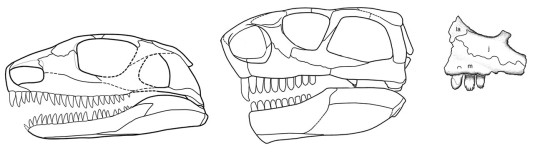
Caseid synapsids are a diverse, long-lived group known from the Late Pennsylvanian–Middle Permian epochs (~300–259 million years ago) of Europe, Russia, and the USA, and, with one exception, all are adapted to eating plants (herbivorous). The most advanced caseids (such as the enormous Cotylorhynchus romeri) have ridiculously small skulls when compared to those of carnivores, spatulate (spoon-shaped) teeth tipped with small tubercles (cuspules) for cropping vegetation, and huge, barrel-shaped ribcages to support a large gut for fermenting cellulose-rich plants. The exception is the earliest known (Late Pennsylvanian epoch, ~300 million years ago) caseid, Eocasea martini, represented by a single, incomplete juvenile specimen from Kansas. The teeth of Eocasea are small and conical, which indicate that it most likely ate insects. Because it’s skull and ribcage are of normal size, in contrast to juveniles of Martenius, Eocasea probably ate insects throughout its life.

Reconstruction of the skull of Martensius bromackerensis (left) from the Early Permian (~290 million years ago) Bromacker quarry, Germany, and the more advanced caseid Ennatosaurus tecton (skull, middle and skull fragment with cuspule-tipped teeth, right), from the Middle Permian (~263 million years ago) of Russia. Skull reconstruction of Martensius made by Diane Scott and modified from Berman et al., 2020. Ennatosaurus skull reconstruction and jaw fragment drawing modified from Maddin et al., 2008.

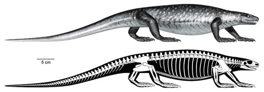
Martensius has a modestly expanded ribcage and a small skull, suggesting that it was herbivorous. Furthermore, the feet of Martensius, like those of other caseids in which the feet are known, are large, with massive, elongated, strongly recurved claws. Martensius also has a well-supported hip region that may have enabled it to rise on its hind legs to reach and tear down overhead branches to feed upon.

The upper and lower teeth of the adult Martensius differ from those of more advanced caseids in being triangular and lacking cuspules. The upper jaw teeth of the juvenile resemble those of the adult, but the lower jaw teeth are more numerous—31 in the juvenile compared to 25 in the adult—and surprisingly, they resemble those of Eocasea. Dave concluded that juveniles of Martensius had teeth adapted for eating insects, which were replaced by an adult dentition that would’ve been good for cropping plants and piercing insects. Remarkably, the juvenile Martensius apparently died while in the process of replacing its juvenile dentition with that of adults.

So why have different juvenile and adult dentitions? Modern animals that eat fibrous plant matter have micro-organisms called fermentative endosymbionts in their large guts, which break down difficult-to-digest plant matter via fermentation. It is assumed that early fossil plant-eaters with broad ribcages also had large guts housing fermentative endosymbionts. Prior to the discovery of Martensius, other scientists hypothesized that early herbivores acquired endosymbionts by eating herbivorous insects that already had these microbes in their guts. In Martensius, the introduction of endosymbionts apparently occurred during the juvenile, insectivorous stage of life, which set the stage for adults to add plants to their diet.

Flesh (top) and skeletal (bottom) reconstructions of Martensius bromackerensis. Illustrations by Andrew McAfee and modified from Berman et al., 2020.

The generic name Martensius honors Thomas Martens for his discovery of vertebrate fossils at the Bromacker quarry and his perseverance in maintaining a highly successful, long-term field operation resulting in the discovery and publication of the exceptionally preserved Bromacker fossils. Bromackerensis refers to the Bromacker quarry, the only locality from which this species is known.

Stay tuned for my next post, which will feature some terrestrial dissorophoid amphibians.

For those of you who would like to learn more about Martensius, here’s a link to the 2020 Annals of Carnegie Museum publication in which it was described.

Amy Henrici is Collection Manager in the Section of Vertebrate Paleontology at Carnegie Museum of Natural History. Museum employees are encouraged to blog about their unique experiences and knowledge gained from working at the museum.

Keep Reading

The Bromacker Fossil Project Part IX: The Dissorophoid Amphibians Tambachia, Rotaryus, and Georgenthalia, Capable Travelers

Filed Under: Blog Tagged With: Amy Henrici, dinosaurs in their time, fossils, Museum from Home, paleontology, Science News, Section of Vertebrate Paleontology, Vertebrate Paleontology

July 29, 2020 by wpengine

Collected on this Day in 1966: Santa Clauses

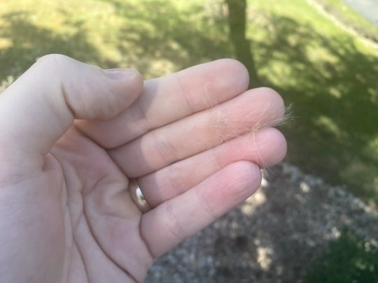
Christmas in July…”Santa Claus” floating in the air.

(Or I guess, technically Boxing Day in July, if that’s a thing.)

thistle seed fluff "Santa Claus" in hand

Make a wish!

Have you ever seen fluff floating by in the air, especially in late July, early August? Kids love chasing the fluff around, often referring to them as “Santas” or “Santa Clauses.”  You catch it, make a wish, and let them go again, floating away.

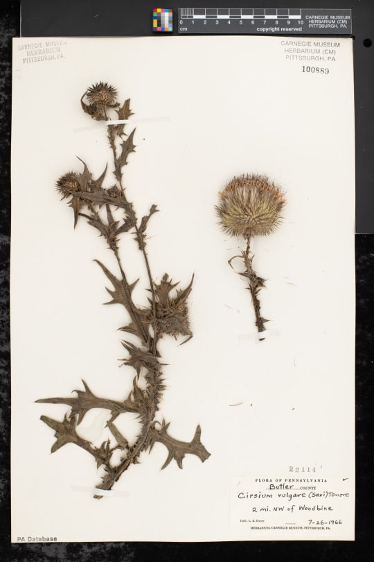
dried thistle specimen on herbarium sheet

These are seeds!  Most likely thistle seeds, like this specimen here.  Or other seeds that have similar “fluff”-like structures.  The botanical term for this “fluff” is pappus.  Pappus is a modified part of flowers in many species in the sunflower family, Asteraceae (think dandelion).  These structures help the seed disperse in the wind, floating away in the breeze, carrying the seed far away.  If you’ve tried to catch them, you know they float away in the air very easily. The seeds are small, and often times have already disconnected from the pappus when you catch them.

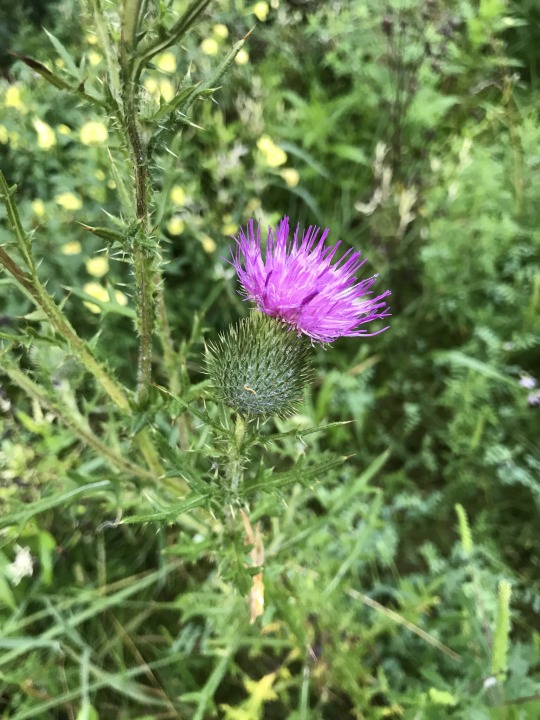
thistle

Check out the “Santa Claus” pappus on this specimen of bull thistle (Cirsium vulgare), collected on July 26, 1966 by Leroy Henry near Woodbine (Butler county), Pennsylvania.  Leroy Henry was a botany curator at the Carnegie Museum.  All species in the genus Cirsium are known as “thistles.”  They have distinctive spiny leaves and stems, with even more distinctive purple flower heads. There are native thistles, but many are introduced. Thistles are common in disturbed areas, and in and around agricultural fields across the country.  Bull thistle is native in Europe and Western Asia, but widely introduced across the world, including North America.  It is the national flower of Scotland, but the species is considered invasive in many places.

Keep an eye out for thistles, and “Santa Clauses.”  Don’t forget to make a wish.

Find this bull thistle specimens here.

Check back for more! Botanists at the Carnegie Museum of Natural History share digital specimens from the herbarium on dates they were collected. They are in the midst of a three-year project to digitize nearly 190,000 plant specimens collected in the region, making images and other data publicly available online. This effort is part of the Mid-Atlantic Megalopolis Project (mamdigitization.org), a network of thirteen herbaria spanning the densely populated urban corridor from Washington, D.C. to New York City to achieve a greater understanding of our urban areas, including the unique industrial and environmental history of the greater Pittsburgh region. This project is made possible by the National Science Foundation under grant no. 1801022.

Mason Heberling is Assistant Curator of Botany at Carnegie Museum of Natural History. Museum employees are encouraged to blog about their unique experiences and knowledge gained from working at the museum.

Related Content

Tulips in June?!

What’s So Special About Poinsettias?

Collected on this Day in 1984: Tobacco

Filed Under: Blog Tagged With: botany hall, collected on this day, Hall of Botany, Mason Heberling, Museum from Home, Science News, Section of Botany

July 20, 2020 by wpengine

A Bumble’s Blog and Bumble “Weee” Catapult Craft

Wander outside in the spring and summer and I bet you will bump into a busy bumble bee bumbling among the wildflowers. Bumble bees (Bombus sp.) are rather rotund, fuzzy bees usually with black and yellow-orange stripes. Unlike the famous honey bee that hails from Europe and Asia, most bumble bee species are native to our area. Bumble bees have small underground colonies with a loose social system, compared to that of a honey bee. While bumble bees produce honey, it is in small quantities and certainly not enough to share on an industrial scale. Still, these fuzzy bumbles play an important role as pollinators of local wildflower populations and are even adapted to pollinate certain flowers!

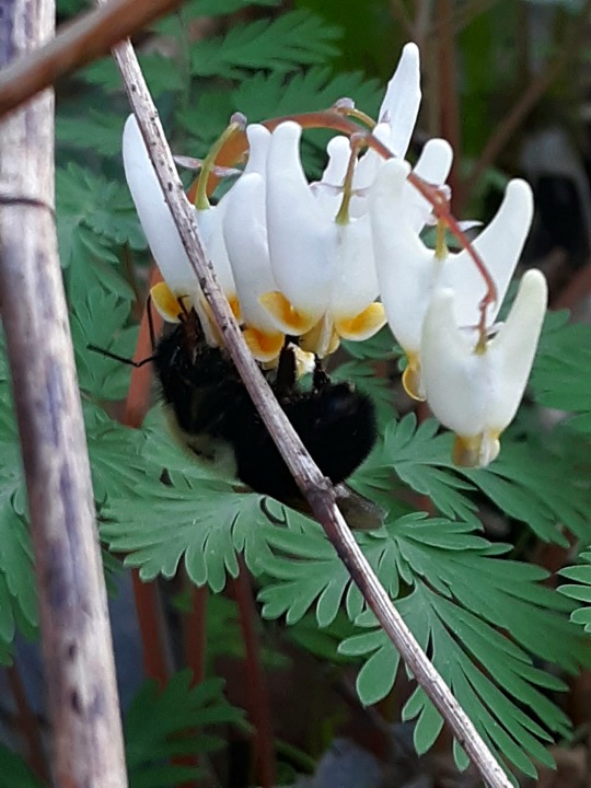
photo of Dutchman's breeches flower

This fashionable bumble bee is trying to squeeze into a little pair of white breeches! Duchman’s breeches (Dicentra cucullaria) to be exact. A bumble bee’s proboscis (tongue) is long enough to reach the nectaries within the nectary spurs or “pant leg” and the bee is strong enough to push open the flower petals to collect pollen.

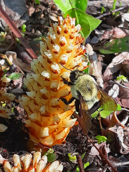
bumblebee on squawroot

This bumble bee is sipping nectar from squawroot (Conopholis americana). Bumble bees and flies are the typical visitors of this parasitic plant! Fun fact: bumble bees “buzz” pollenate, which means they vibrate their bodies, effectively knocking pollen down into the flowers they visit.

Upon observing the flight of a bumble bee, I have noticed that while they are strong flyers, they are not the most graceful. Sometimes they miss their mark and land on a chunk of moss instead of the flower. However, they just take that moment to rest their wings before firing up their little motor and buzzing off into, hopefully, the next flower. Their rather clunky flight pattern gave me an idea for a fun activity to help young children learn about pollinators (and sneak in a STEM activity): The Bumble “Weee” Catapult! See below for assembly instructions:

Materials

      3 pipe cleaners

      Paper

      Pen or pencil

      Coloring supplies

      Scissors

      Recycled egg carton

      Recycled plastic spoon

      4-5 large Rubber bands

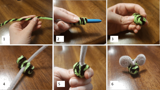
Let’s dismiss the idea of launching real bumble bees and begin building the bee puppet 😉. Pick 3 different colors of pipe cleaner. Feel free to go with traditional bee colors or mix it up!

1.     For the body, twist together two pipe cleaners.

2.     Wrap the twisted pipe cleaners tightly around a pen or pencil and slide it off.

3.     Tuck the loose ends inward and tighten up the coils on the end you would call the head.

4.     For the wings, shorten the remaining pipe cleaner by 1/3, then loop it under one of the coils of the bee’s body.

5.     Adjust the pipe cleaner for equal length on either side and twist at the base.

6.     Roll in the loose ends to finish forming your wings, thus completing the bumble bee.

step by step photos of creating a bee from pipe cleaners

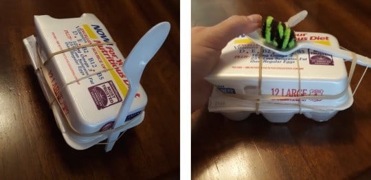
Next, build the catapult to help your bee puppet fly. The catapult consists of half of an egg carton, 4-5 rubber bands, and a recycled spoon. Tension energy is generated when the spoon is pulled back. The arm stores that energy as potential energy. Upon release, that potential energy is transferred to the object as kinetic energy, moving the object away from the arm. Pictured is the simplest catapult design with rubber bands holding the spoon, or arm of the catapult, in place. Feel free to design something more elaborate!

photo of catapult made from egg carton, plastic spoon, and rubber bands

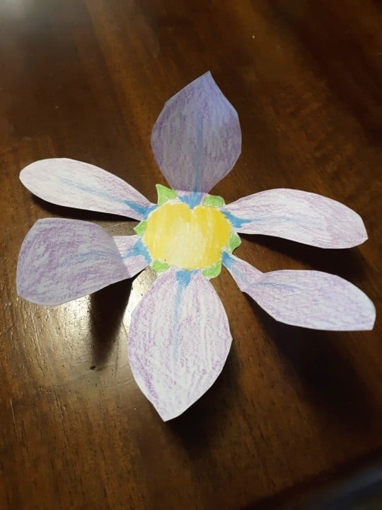
Finally, the bumble bee needs a flower to land in! Draw a flower of your choice on a piece of paper and color it in. Many bees are able to see UV light, which means they are able see colors and patterns invisible to the human naked eye. Some flowers have nectar guides that really stand out to bees, so feel free to draw some nectar guides, or lines that point to the center on your flower targets to help guide your bumble bee!

photo of paper flower

Feel free to create as many flowers as you like and propel your little bumble bee into as many flowers as you can. Happy pollinating!

Sara Klingensmith an educator at Powdermill Nature Reserve, Carnegie Museum of Natural History’s environmental research center. Museum employees are encouraged to blog about their unique experiences and knowledge gained from working at the museum.

Related Content

Jumping Spiders with Sebastian

Pittsburgh Dinosaur Driving Tour

Photogenic Fungi

Filed Under: Blog Tagged With: activities, Crafts, Museum from Home, Nature 360, Nature Lab, Powdermill, Powdermill Nature Reserve, Sara Klingensmith

July 15, 2020 by wpengine

The Bromacker Fossil Project Part VII: Eudibamus cursoris, the Original Two-legged Runner

New to this series? Read The Bromacker Fossil Project Part I, Part II, Part III, Part IV, Part V, and Part VI. 
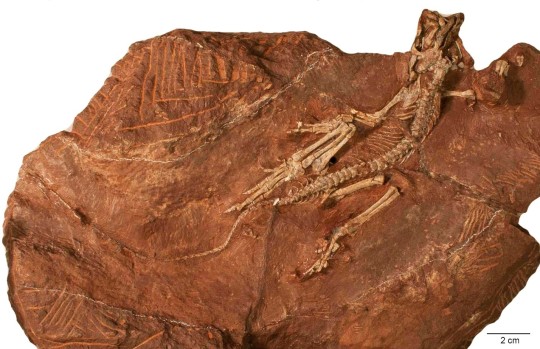
Holotype specimen of Eudibamus cursoris, the most complete bolosaurid reptile known. Photo by the author, 2013.

Stuart Sumida discovered some small bones in the Bromacker quarry in 1993, the same year that the holotype skeleton of Diadectes absitus was found. Dave Berman told me that when Stuart showed them to him, he couldn’t see anything because they were so small. Upon closer examination, Dave, Stuart, and Thomas Martens identified them as those of the captorhinomorph reptile Thuringothyris mahlendorffae. Thomas’ wife Stefani, whose maiden name is Mahlendorff, discovered the first specimen in the Bromacker in 1982, and Thomas and a colleague named it in her honor in a 1991 publication.

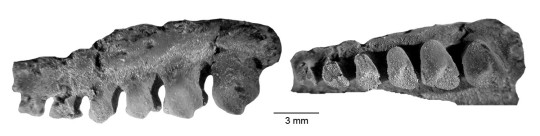
The fossil was exposed in several pieces of rock, which Thomas shipped to Carnegie Museum of Natural History (CMNH) along with the large block of rock containing Diadectes. I didn’t prepare the specimen until several years later, as other projects, including the Diadectes, overshadowed it. Once I began working on it, though, Dave and I realized that it was not Thuringothyris. Indeed, we had no idea what type of animal it was, and our puzzlement grew as I exposed more of it. The identity wasn’t revealed until I had uncovered some very unusual, tiny teeth, which under the high magnification of the preparation microscope appeared to have a bulbous cusp towering over a basin. They looked vaguely familiar to me, but because I couldn’t immediately put a name on them, I rushed to get Dave from his office. Once Dave saw the teeth, he realized that the specimen was a new genus and species in the rare, enigmatic reptile group Bolosauridae.

Tiny teeth of a bolosaurid reptile, Bolosaurus striatus, in lateral (side; left) and occlusal (chewing surface; right) views. The specimen is in the CMNH Vertebrate Paleontology collection. Photos by Spencer Lucas (Research Associate, CMNH).

Until the discovery of Eudibamus cursoris, bolosaurids were represented in the fossil record by two genera, Bolosaurus and Belebey, which were based mainly on poorly preserved skull and fragmentary jaw fossils from Texas and Russia, respectively. Even though bolosaurids had been known since 1878, their relationship to other reptiles was not well understood. The nearly complete anatomy of Eudibamus allowed our team to determine that bolosaurids are the oldest member of the ancient group of reptiles called Parareptilia. This group has no living relatives, except possibly for turtles, a hypothesis that is highly debated by scientists.

Eudibamus cursoris fossil
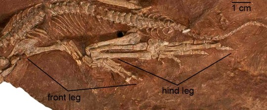
Closeup of front and hind legs of Eudibamus. The hind leg, folded upon itself, is considerably longer than the front leg. Photo by the author, 2013.

When our study of the fossil began, we realized that Eudibamus was very different than other reptiles from that time. Proportions of the limbs and positions of the articulation surfaces on the upper and lower hind leg bones indicated that, in terms of posture, Eudibamus resembled a bow-legged human with a bad back instead of a typical sprawling reptile on four legs. It could stand and locomote on its hind legs in an upright posture (bipedal) with its legs held close together and in the same plane (parasagittal).

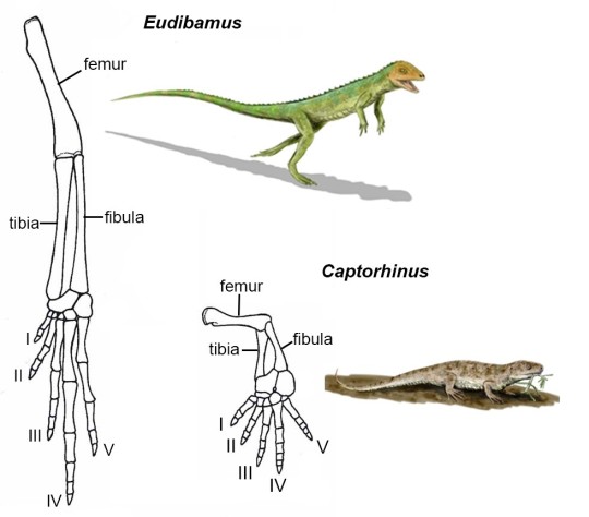
Dave was in constant phone communication with team member Dr. Robert Reisz (Professor, University of Toronto at Mississauga). One day Robert called Dave to ask if all the tail had been exposed, because he learned that modern lizards that are able to run bipedally have a long tail to help maintain their balance. The specimen was in Dave’s office and he immediately uncovered more of the tail and then let me finish the task. The tail was indeed very long and extended close to the edge of the block, which I had previously reduced in size. Additionally, we determined that the third, fourth, and fifth toes of the hind foot also were greatly elongated through lengthening of some of the individual toe bones, and that the first and second toes were extremely shortened by the reduction in size of individual toe bones. We hypothesized that when Eudibamus ran bipedally, it would rise on its toes, so that only the tips of the third, fourth, and fifth toes would contact the ground.

Drawing of the hind leg of Eudibamus cursoris (left) and the roughly contemporaneous reptile Captorhinus (right). Leg drawings are scaled to the same torso length of the whole animal. Illustrations of the animals are not to scale. Hind leg drawings are modified from Berman et al., 2000 and animal illustrations are from Wikimedia Commons.

Eudibamus occurred at least 60 million years before other bipedal, parasagittally-running reptiles appeared in the fossil record. This is reflected in its scientific name, which is derived from the Greek “eu,” meaning original or primitive, and “dibamos,” meaning on two legs. “Cursoris” is Latin, meaning runner. Examples of other reptiles using this locomotion mode are the dinosaurs Allosaurus fragilis and Tyrannosaurus rex, which you can view in CMNH’s Dinosaurs in Their Time exhibition.

So, what was the advantage of being able to run bipedally instead of running on all four legs? Lengthening the hind leg and foot would greatly increase stride length, especially if only the tips of the toes contacted the ground, which is an efficient way to increase speed. Eliminating arm to ground contact while running removes forelimbs from the path of the long-striding hind legs. The bulbous teeth and jaw structure of Eudibamusindicate that it was herbivorous. It seems likely, then, that Eudibamus used its ability to sprint to avoid becoming a tasty meal for a pursuing predator.

Eudibamus cursoris illustration
Peter Mildner (exhibit preparator at the Museum der Natur, Gotha) made a surprise visit to the Bromacker one afternoon to show us a model of Eudibamus cursoris he’d made. This image shows the model in the present day Bromacker quarry, part of the region it inhabited 290 million years ago. Photo by the author, 2006.

One of our laments is that a fossil trackway preserving Eudibamus walking quadrupedally and then switching to a bipedal gait has yet to be found.

Next time you are at CMNH, make sure you see the cast of the fossil skeleton and a model of Eudibamus that are exhibited in the Fossil Frontiers display case in CMNH’s Dinosaurs in Their Time exhibition. Stay tuned for my next post, which will feature the herbivorous mammal-like reptile Martensius bromackerensis.

For those of you who would like to learn more about Eudibamus, here is a link to the 2000 Science publication in which it was described: https://science.sciencemag.org/content/290/5493/969.

Amy Henrici is Collection Manager in the Section of Vertebrate Paleontology at Carnegie Museum of Natural History. Museum employees are encouraged to blog about their unique experiences and knowledge gained from working at the museum.

Keep Reading

The Bromacker Fossil Project Part VIII: Martensius bromackerensis, Honoring a Colleague

Filed Under: Blog Tagged With: Amy Henrici, dinosaurs in their time, Museum from Home, Science News, The Bromacker Fossil Project, Vertebrate Paleontology

  • « Go to Previous Page
  • Page 1
  • Interim pages omitted …
  • Page 11
  • Page 12
  • Page 13
  • Page 14
  • Page 15
  • Interim pages omitted …
  • Page 26
  • Go to Next Page »

sidebar

About

  • Mission & Commitments
  • Directors Team
  • Museum History

Get Involved

  • Volunteer
  • Membership
  • Carnegie Discoverers
  • Donate
  • Employment
  • Events

Bring a Group

  • Groups of 10 or More
  • Birthday Parties at the Museum
  • Field Trips

Powdermill

  • Powdermill Nature Reserve
  • Powdermill Field Trips
  • Powdermill Staff
  • Research at Powdermill

More Information

  • Image Permission Requests
  • Science Stories
  • Accessibility
  • Shopping Cart
  • Contact
  • Visitor Policies
Stay in the loop! Sign up for our newsletter(s).
One of the Four Carnegie Museums | © Carnegie Institute | Privacy Policy | Terms of Use | Accessibility
Rad works here logo