• Skip to primary navigation
  • Skip to main content
  • Skip to primary sidebar

Carnegie Museum of Natural History

One of the Four Carnegie Museums of Pittsburgh

  • Visit
    • Buy Tickets
    • Visitor Information
    • Exhibitions
    • Events
    • Dining at the Museum
    • Celebrate at the Museum
    • Powdermill Nature Reserve
    • Event Venue Rental
  • Learn
    • Field Trips
    • Educator Information
    • Programs at the Museum
    • Bring the Museum to You
    • Guided Programs FAQ
    • Programs Online
    • Climate and Rural Systems Partnership
  • Research
    • Scientific Sections
    • Science Stories
    • Science Videos
    • Senior Science & Research Staff
    • Museum Library
    • Science Seminars
    • Scientific Publications
    • Specimen and Artifact Identification
  • About
    • Mission & Commitments
    • Directors Team
    • Museum History
  • Tickets
  • Give
  • Shop

Blogs from our Scientific Researchers

Carnegie Museum of Natural History is home to active research and vast scientific collections. Our scientific researchers regularly contribute to the blog at the museum.

January 21, 2021 by wpengine

A Head Above the Rest: Unearthing the Story of Our Leatherback Sea Turtle

When you think of BIG sea creatures, you probably imagine great white sharks, huge blue whales, or ginormous cephalopods like the giant squid (or, for the more imaginative, the Kraken!). But would you believe me if I told you that the ocean is also home to a reptile that grows far larger than a human? Many people are familiar with the “typical” green sea turtle (Chelonia mydas) or even the hawksbill sea turtle (Eretmochelys imbricata), known for its beautifully patterned shell. However, these species are dwarfed in size compared to the leatherback sea turtle (Dermochelys coriacea). Adult leatherback sea turtles are usually 6 to 8 feet long and 550 to 1500 pounds. To put that into context, imagine 3 to 8 adult men of average height and weight huddled together or 8 to 24 Labrador retriever dogs playing about in a group (now THAT would be heavenly!). An animal that big takes up a great deal of room, which is fine in the expansive ocean but is rather problematic if such a turtle is to become a museum research specimen. That is exactly the case with CM 44460, the famous leatherback sea turtle housed in the Carnegie Museum of Natural History’s Section of Amphibians and Reptiles.

Head and esophagus of leatherback sea turtle CM 44460. The esophagus is so large it needed to be split into pieces— the two circles at the lower left and right corners of the tank and the large mass in the top left corner. Leatherback sea turtles lack teeth, and instead rely on spikey protrusions present in their mouths and esophagi to keep down their favorite prey item, jellyfish.

Cast of CM 44460 hanging above visitors in Discovery Basecamp.

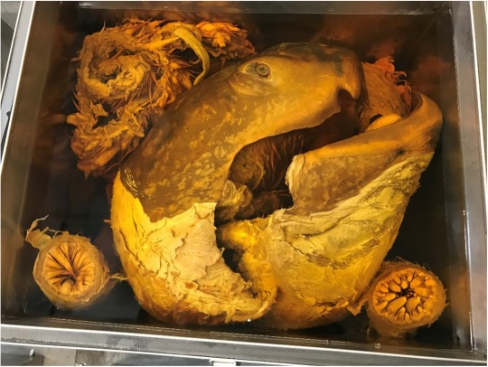
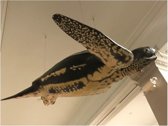
When people tour the Section of Amphibians and Reptiles, we make it a point to open one of the first metal tanks our guests see, tank 156. This tank houses a single impressive specimen – the giant head and esophagus of a leatherback sea turtle. I started as the collection manager of the section just under two years ago and, until recently, the only information I had for this specimen were the scant details noted in the section’s database and on a printed sheet attached to the lid of the tank: a fisherman had found the specimen dead when it washed ashore in Maine in 1965. That was it. I knew the entire animal (not just the head) had washed ashore since a cast was made of the body and that replica is now hanging in Discovery Basecamp. I also knew ecological and herpetological information about the species in general, but nearly every specimen in a natural history collection has a story, and I knew this one had to be good… but I didn’t know what it was…

… until I began digitizing the section’s archives.

Let’s take a step away from our leatherback sea turtle specimen to understand what “digitizing the section’s archives” really means. Carnegie Museum of Natural History is over 100 years old, and the herpetology archives date back to the museum’s inception. That means we had, at the time I became involved with the digitizing work, nearly 125 years of correspondence, field notes, specimen data, and collection-related events to clean, scan, and properly organize and house both physically and electronically. (For a more in-depth dive into this archiving process, see section archivist Ren Jordan’s post here.) It took a team of about 10 people (part-time and full-time interns, work-study students, and staff members) over a year to complete this daunting task. The treasure trove of information we unearthed in those archives is priceless, and CM 44460’s story is a treasure worth sharing.

Images from archives showing how staff members prepared CM 44460 to be accessioned into the herpetology collection and displayed to the public. Clockwise from the top left: Herpetology staff members C. J. McCoy and Arthur Bianculli lift the shell onto a cart for transport; Herpetology curator Neil D. Richmond and museum preparator Otto M. Epping measure out the cast of CM 44460 created from the shell and body measurements; Preparator Otto M. Epping and Exhibits staff member Forest Hart removing the shell from a cargo van upon arrival to the Carnegie Museum; Herpetology staff members C. J. McCoy, Arthur Bianculli, and Neil D. Richmond examine the head of CM 44460 in a large potato chip can; Herpetology curator Neil D. Richmond shows the head to museum director M. Graham Netting as another staff member looks on.
black and white photo of three men pulling a turtle head out of a can

Herpetology staff members C. J. McCoy, Arthur Bianculli, and Neil D. Richmond examine the head of CM 44460 in a large potato chip can upon its arrival to the museum (A). The complete description of the image as it appears affixed to the back of the image (B).

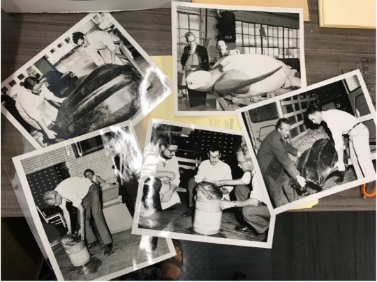
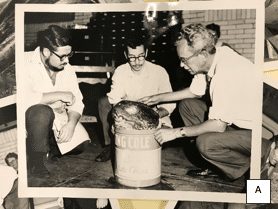
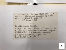
During the digitization work, the archival material I processed included the field notes of past-curator Dr. C. J. McCoy, and among his papers was a crumbly old folder labeled “CM 44460” that required rehousing. The number lacked any context for me at the time because the section has over 180,000 catalog (or CM) numbers and, try as I might, I don’t yet have them all memorized. When I pulled out pictures from the folder, though, CM 44460’s identity instantly became apparent, for I found myself looking at the images of our famous leatherback sea turtle. One picture showed the creation of the cast and another depicted the shell being carried by two men due to its size. Another image showed Dr. McCoy crouched with two other men near a huge open tin can labeled “Potato Chips” with, shockingly, the head of dear CM 44460 peeking out of the top. A note affixed to the back of the image read “C. J. McCoy, Arthur Bianculli, & Neil D. Richmond examining head which filled 7-gal. can. 27 Aug. 1965. Leatherback Turtle caught 16 Aug. 1965 off Swan’s Is., Maine by Lobsterman Robert Joyce. Presented to Carnegie Museum by Dave Shelton, Aqualand, Bar Harbor, Maine” (Image 4B). Suddenly pieces of the story were falling into place. This specimen was transported from Maine to Pittsburgh in pieces, with the head arriving separate from the body and shell in a 7-gallon potato chip container!

A couple months later, I unearthed another folder in the archives with data from the specimen. The documents recorded the preservation process of the turtle, including measuring and weighing different organs (knowing that they would be too large to properly preserve and store), and how long it took the head to become fully and properly fixed in formalin. Through these notes, I learned that the turtle was a female measuring 7’5” from the tip of her tail to the tip of her snout, and that her ocean wandering was powered by a flipper-span of 8’4”! Based upon her carapace (the top part of a turtle shell) measuring in at 5’5”, this turtle was likely sexually reproductive and, therefore, rather old. CM 44460’s story is so much clearer now and really goes to show how each specimen in a collection has its own unique history just waiting to be investigated.

Stevie Kennedy-Gold is the collection manager for the Section of Amphibians and Reptiles at Carnegie Museum of Natural History. Museum employees are encouraged to blog about their unique experiences and knowledge gained from working at the museum.

Related Content

Ask a Scientist: What is a pitfall trap (for herpetology)?

Do Snakes Believe in the Tooth Fairy?

Lantern Slides

Filed Under: Blog Tagged With: amphibians and reptiles, herpetology, Museum from Home, Science News, Stevie Kennedy-Gold

January 19, 2021 by wpengine

Insect metamorphosis: the key to a fresh new start

For many people, the new year represents an opportunity to make a fresh start, consider self-improvement, or turn over a new leaf. As in all fields of human endeavor, insects are way ahead of us and have already developed the ultimate technology for personal reinvention: metamorphosis.

drawing of the stages of metamorphosis

Among entomologists, “metamorphosis” refers to the process by which a tiny hatchling insect becomes a fully functioning adult. This process can take place in two ways. Incomplete metamorphosis is the process by which an insect molts through a series of increasingly large, adult-like stages (“instars”) before completing the final molt into an adult. Insects that develop this way include grasshoppers, stink bugs, dragonflies, termites, and mantises.

drawing of various insects including a butterfly, bee, and beetle


Complete metamorphosis, on the other hand, involves a (typically) worm-like larva which undergoes a quiescent, or inactive, pupal stage before reaching adulthood. Insects that undergo complete metamorphosis include beetles, ants, bees, wasps, lacewings and antlions, flies, and moths. These orders are often described as “holometabolous,” which simply means that their development includes pupation.

drawing of a moth teaching other moths about cocoons and turning to "mystery goo"


The process of pupation is fascinating and mysterious: essentially, the caterpillar zips itself up into a sleeping bag made of its own skin, turns to soup, and comes out a butterfly. How?

In fact, insect pupation remained a scientific mystery for many years, largely because of the difficulty in observing the pupation process without destroying or interfering with development. However, interfering with development turned out to be the key to understanding this process: early investigators (e.g. Jan Swammerdam, the 17th century microscopist) discovered that structures corresponding to the approximate positions of future wings could be dissected from within late stage, prepupal larvae. Several centuries later, the ability to induce fluorescence in selected cell lines allowed researchers to observe the activity of these future wings, legs, and antennae throughout larval development. This research led to the identification of what are now known as “imaginal discs.”

caterpillar wearing headphones holding a record called "I, Ron Butterfly"

Here’s how it works: secret little collections of cells are formed during embryogenesis, and rest dormant inside the larva as it grows. The larva and its essential larval structures (usually the digestive system) grow larger, but the dormant cells do very little. These cells are known as imaginal cells and their aggregate structures are called imaginal discs (The term refers not to imagination, but to the imago, a synonym for the insect’s adult stage). The cells within these imaginal discs are largely dormant until a special cue— temperature, day length, growth, or otherwise— triggers the hormones that kickstart pupation. The larva forms a tough outer casing from its outermost exoskeleton or uses silk glands to create a protective nest (e.g. a cocoon).

metamorphosis diagram
Image source: Aldaz, S. and Escudero, L.M., 2010. Imaginal discs. Current Biology, 20(10), pp.R429-R431.

As pupation begins and the larval body breaks down into fluid, the imaginal discs begin to undergo rapid development, telescoping outward to form the longer legs, wings, antennae, mouthparts, and other complex adult body structures. The only remnants of the larva that stay functional are the tracheae, hollow tubes which allow it to breathe.

Once the adult structures are fully formed, they will remain soft in order to fit inside the now too-small pupa. The pupal case splits open, and the newly emerged adult insect forces air and fluid into its new wings to unfurl them fully before they harden.

butterfly emerging from cocoon
Image from Creative Commons.

Forming a hard outer casing and liquefying your existing body may not sound like an inspirational concept for the new year, but perhaps it should. The lesson of the butterfly is that the developmental foundations of the beautiful, functional adult were inside the awkward, squirmy larva all along. The imaginal cells were always there, just waiting to be awakened.

For more discussion of insect pupation and tips on using caterpillars to get kids into science, see this previous IZ blog post by Dr. Jim Fetzner, “Kids and Caterpillars: Fostering a Child’s Interest in Nature by Rearing Lepidoptera (Moth and Butterfly) Larvae.”

Ainsley Seago is Associate Curator of Invertebrate Zoology. Museum employees are encouraged to blog about their unique experiences and knowledge gained from working at the museum.

Related Content

O-Do-Nates or O-Don’t-Nates: Dragonflies and Damselflies in the Section of IZ

Looking for Bugs in Your Yard

Why One Bug Just Won’t Do…

Filed Under: Blog Tagged With: Ainsley Seago, insects, Invertebrate Zoology, Museum from Home, Science News

January 11, 2021 by wpengine

2020 Rector Christmas Bird Count Results

Black-capped Chickadee

On December 20, 2020, 34 intrepid birders braved a wintry mix to count birds in assigned sectors within a 15-mile diameter circle centered just northwest of Powdermill Nature Reserve. Another eight, who lived within designated territory, closely watched their bird feeders and yards for avian visitors. Why would so many birders be out in less-than-ideal weather conditions? They were all participating in the Rector Christmas Bird Count.

Map of the Rector Christmas Bird Count circle with each count sector outlined with red, created by James Whitacre, GIS Research Scientist at Powdermill Nature Reserve.

Christmas Bird Count (CBC) is an annual event sponsored by the National Audubon Society that happens in mid-December through early January, with the compiler of each count circle choosing a specific count date within that timeframe. This year marks the 121st anniversary of the activity. The count started on Christmas day in 1900 with the purpose of censusing birds by counting them in the field using optics rather than by using shotguns. Although there were only 25 count circles in the first CBC, it has grown into an international event with nearly 2900 circles spread across the Western Hemisphere and even to Pacific Islands as far away as Guam and the Northern Mariana Islands.

Today, the CBC is a fun day for birders and bird watchers of all skill levels to head outside with the goal of identifying and counting every bird they see and hear within their count areas. The data gathered though this citizen science initiative contributes to both long-term and short-term population studies. To date, more than 200 peer-reviewed scientific publications have used CBC data in their analyses.

Although the count was a bit different this year with COVID-19 precautions keeping counters in different germ pools separate, we had an excellent turnout of both advanced and beginner birders, including some young birders.

Eastern Screech Owl
Red-headed Woodpecker

And what a count it was! This winter is an irruption year (for more info on irruptive migration, please see this blog.) for many species, and although we didn’t find hordes of these birds during the count, we did see Pine Siskins, Purple Finches, Red-breasted Nuthatches, higher-than-average numbers of Black-capped Chickadees, and the much sought after Evening Grosbeak. The counters recorded many interesting and less common species this year, including the count’s third ever Snow Goose, third ever Eastern Phoebe, and fifth ever Common Yellowthroat. Both the phoebe and Common Yellowthroat are species that winter in the southeastern US. Counting efforts that began an hour before dawn produced exceptional owl numbers (eight Eastern Screech-Owls, one Great Horned Owl, and two Barred Owls). Additionally, the birders recorded high counts for several species including Ruddy Duck, Black Vulture, Bald Eagle, Red-shouldered Hawk, Red-bellied Woodpecker, Common Raven, Carolina Wren, and Song Sparrow. Most notably, the group counted a record-setting seven Red-headed Woodpeckers, a species that is uncommon in southwestern Pennsylvania and can be reliably found in only one spot of suitable habitat within the count circle.

Carolina Wren

We thank all of the participants for a wonderful count this year! In all, we tallied 4361 birds of 69 species, a remarkable result thanks to the valiant effort of all of the counters. We look forward to hosting the Rector count next year!

Annie Lindsay is the Bird Banding Program Manager at Carnegie Museum of Natural History’s Powdermill Nature Reserve. Museum employees are encouraged to blog about their unique experiences and knowledge gained from working at the museum.

Related Content

Ask a Scientist: Ravens and crows, what’s the difference?

Oh deer, that’s a lot of parasites!

Snowflakes and Snow Fleas

Filed Under: Blog Tagged With: Annie Lindsay, Birds, Powdermill Nature Reserve, Science News

January 8, 2021 by wpengine

A Visit to the Mammoth Site, Hot Springs, SD

Did you know that not all museums display their fossil specimens mounted in life-like poses? At The Mammoth Site of Hot Springs, South Dakota, visitors view fossils “in situ,” or as they were discovered, and because excavation continues year-round, this unique museum is also an active dig site.

brown sign that says The Mammoth Site
A sign welcomes visitors to The Mammoth Site of Hot Springs, South Dakota.

Instead of being held in place by the fabricated support structures that are so crucial to traditional fossil displays, bones at The Mammoth Site rest on sediment and appear in the same orientations in which they were found. The remains of more than 60 Columbian mammoths (Mammuthus columbi) have been documented here to date, comprising the world’s most extensive collection of skeletons of these Ice Age elephant relatives.

The Mammoth Site was discovered in 1974 when the landowner decided to build a housing development on the 14-acre plot. While the heavy machine operator was bulldozing a small hilltop, he found tusks and bone. Construction stopped and officials at four colleges were contacted, but none expressed interest in the find. Fortunately, the son of the heavy machine operator, who had taken geology and archaeology courses in college, was able to gain interest from one of his former professors, who was then conducting fieldwork in Arizona. When the professor arrived at the site a few days later he recognized the exposed bones of four to six individual mammoths and the potential for more nearby. He arranged for a field crew to salvage and stabilize the visible bones, teeth, and tusks, and returned the next summer with a group of students to do more excavating. A complete skull with tusks attached was the prize find of these more organized recovery efforts, and by the end of the summer the landowner had decided the tract’s highest value was as a place for scientific study.

mammoth skulls in situ
Mammoth skulls with tusks attached at The Mammoth Site. Notice the sediment supporting the fossils.

I recently had the opportunity to visit The Mammoth Site, which is located in the Black Hills, a scenic region of green pine trees and deep red earth. Once you purchase your admission, you are directed to a theater where a looped video introduces the relevant geologic history. The site is the result of a sinkhole that developed when groundwater dissolved the limestone layers through which it flowed. Subterranean water-filled caverns were an early product of this process, but as the water table lowered the caverns weakened and collapsed, resulting in a deep sinkhole with a chimney-like shaft, through which a warm artesian spring percolated to the surface. In three phases over a period of 750 years, the sinkhole refilled with sediment and the remains of mammoths and other creatures before it was eventually reduced to a mud wallow.

photo of geologic map
Geologic map of the beautiful Black Hills area of South Dakota and Wyoming.

After the theater, the bonebed is the next stop. The museum has a special app that can be listened to with headphones for a tour of the bonebed. The bonebed room is very large and naturally lit and has a high beam ceiling with windows at the top of one wall. There is a crane attached to the rafters that is used to move any specimens that need to be permanently removed from the ground. Because a large tusk can weigh over 100 pounds, and skulls far more than this, this overhead crane is an essential tool.

complete mammoth skeleton
The most complete mammoth skeleton or “model mammoth,” found in the deep end of the bonebed. It is used to compare to the remains of others to determine attributes such as age, size, and sex.

How, you ask, do researchers know there are over 60 individuals in the sinkhole? For every mammoth or person or other critter with a skeleton, there are a certain number of each bone in the body. Because mammoths have two tusks it is possible to count the number of tusks in the bonebed, 123, and divide by two to calculate the presence of at least 62 individuals.

bonebed at Mammoth Site
How many tusks can you find in this section of the bonebed?

Determining the sex of a mammoth is possible when its pelvis is well-preserved with minimal crushing or distortion. By measuring a specific spot on the pelvis and the width of the pelvic canal at a certain area, and comparing these two measurements, it can be determined whether the pelvis belonged to a male or female mammoth. This calculation is possible because males are generally larger than females, and also because females had a proportionally larger pelvic canal to aid in giving birth. Mammoth remains recovered at The Mammoth Site have all been male. Although the presence of more than 60 males but no females at the site may seem surprising, studies have shown that “natural death traps” such as The Mammoth Site captured many more males than females. This may be because, rather than living in herds led by a knowledgeable matriarch, relatively inexperienced male mammoths typically traveled alone, making them more likely to get stuck in these kinds of traps.

It is also possible to age a mammoth using growth rates of bones and the state of fusion of the epiphyses (the ends of the limb bones); however, it is most accurate to age these animals by measuring their teeth. The length and width of the occlusal (= chewing) surface is then used to verify which of their six sets of teeth they were using at the time of death. Generally, a mammoth’s life span could be as long as 60 to 80 years, an age when the animal would be relying upon its sixth set of teeth. When these teeth wore down, starvation would follow. Dental comparisons at The Mammoth Site indicate that most of the remains represent mammoths that were between 15 and 29 years old when they died, with a few in their late forties or early fifties.

mammoth skull fossil in situ
An upside-down mammoth skull shows holes at the front where the tusks attach. Two sets of molars are also visible (I think).

When you next visit Carnegie Museum of Natural History, be sure to head to Pleistocene Hall, where we have our very own mounted Columbian mammoth skeleton on display!

mounted mammoth fossil
The mounted Columbian mammoth at Carnegie Museum of Natural History.
And please remember to keep a tusk-length apart! (Social distancing the mammoth way.)

Linsly Church is a Curatorial Assistant in the Section of Vertebrate Paleontology at Carnegie Museum of Natural History. Museum staff, volunteers, and interns are encouraged to blog about their unique experiences and knowledge gained from working at the museum.

Related Content

Ask a Scientist: What kind of dinosaur is a megaraptorid?

Early Bats: Ancient Origins of a Halloween Icon

Mesozoic Monthly: Vegavis

Filed Under: Blog Tagged With: Linsly Church, Museum from Home, Science News, Vertebrate Paleontology

January 7, 2021 by wpengine

Mesozoic Monthly: Vegavis

Disclaimer: Our dinosaur paleontologist Matt Lamanna typically edits Lindsay Kastroll’s Mesozoic Monthly posts before they go live, but due to some much-needed holiday revelry he was late in getting to this one. As such, it’s being posted in January rather than in December as Lindsay had intended. Matt sends his apologies!

‘Tis the season for eating candy canes, singing Christmas carols, and kicking off a new year of Mesozoic Monthly! That’s right – one year ago, the first Mesozoic Monthly debuted in December 2019, spotlighting the ceratopsian dinosaur with a candy cane-shaped nasal horn, Einiosaurus. This December, we’ll move from candy canes to carols as we feature Vegavis iaai, the first Mesozoic bird known to have had a syrinx (the avian “voice box”)!

Photo (left) and computed tomographic (CT) scan image (right) of the type, or name-bearing, specimen of Vegavis iaai, a partial skeleton inside a ~70-million-year-old rock concretion from Vega Island, Antarctica. Photo from the Antarctic Peninsula Paleontology Project website.

Birds evolved during the Mesozoic Era, the so-called “Age of Dinosaurs,” before non-avian dinosaurs became extinct. Last month, for the November edition of Mesozoic Monthly, we discussed what makes modern birds members of the group of theropod dinosaurs, but what I didn’t mention is that birds lived alongside non-avian dinosaurs! Birds evolved around 165 to 150 million years ago during the Jurassic Period, the second of three time periods in the Mesozoic. The Jurassic dinosaur Archaeopteryx represents a transitional stage between birds and non-avian dinosaurs: its fossils display obvious flight feathers like a bird, but it also has many non-avian dinosaur characteristics such as a toothy mouth, a long bony tail, and even a miniature version of a killing claw like that of Velociraptor.

Replica skeleton of Archaeopteryx lithographica on display here at CMNH. Photo from Wikimedia Commons.

Birds lived and evolved alongside their non-avian relatives for almost 100 million years, and by the end of the Cretaceous Period (the third and final time period of the Mesozoic), the distinct groups of birds that we recognize today were beginning to originate. Vegavis was an ancient relative of ducks and geese discovered on Vega Island, an island off the coast of the Antarctic Peninsula (the part of Antarctica that juts northward towards South America). At that time, Antarctica was warmer than it is now and home to lush temperate forests.

Sandwich Bluff, the site on Vega Island, Antarctica that has produced all known fossils of Vegavis. Photo by Eric Roberts, James Cook University.

With many skeletal features suggesting that it was a diving bird that propelled itself with its feet, Vegavis was probably as well-adapted to life in the water as it was to life in the skies. While it’s certainly incredible that scientists are able to deduce this much information about its behavior from just its skeleton, the story gets better: one specimen of Vegavis includes a fossilized syrinx, the organ that birds use to produce sound! A syrinx’s shape is directly related to the sounds it can make, and the fossilized syrinx of Vegavis was a distinctively goose-like asymmetrical shape. So, this ancient bird may well have honked! If it did, it would have sounded much more like six geese-a-laying than, say, four calling birds, three French hens, two turtle doves, or a partridge in a pear tree.

Lindsay Kastroll is a volunteer and paleontology student working in the Section of Vertebrate Paleontology at Carnegie Museum of Natural History. Museum staff, volunteers, and interns are encouraged to blog about their unique experiences and knowledge gained from working at the museum.

Related Content

Ask a Scientist: What kind of dinosaur is a megaraptorid?

The Bromacker Fossil Project XIII: What We Learned

Pebble or Jaw?

Filed Under: Blog Tagged With: dinosaurs in their time, Lindsay Kastroll, Mesozoic Monthly, Science News, Vertebrate Paleontology

December 30, 2020 by wpengine

The Bromacker Fossil Project Part XIII: What We Learned

New to this series? Need to catch up on your reading? Here are all the previous posts for the Bromacker Fossil Project: Part I, Part II, Part III, Part IV, Part V, Part VI, Part VII, Part VIII, Part IX, Part X, Part XI, and Part XII. 

Collage of the fossils highlighted in this series. Images not to scale. Photos by the author, Dave Berman, and Thomas Martens.

The Bromacker quarry is a rare site in that it preserves exquisite, articulated fossils of a unique vertebrate fauna that lived in an atypical or rarely recorded Early Permian (~290 million years ago) setting. Early in our work at the Bromacker, we became aware that the fossil vertebrates we were finding were unknown or extremely rare in Europe but were closely related or identical to species commonly found in North America. Until then, most of the fossil vertebrates found in Europe were discovered in gray to black sediments deposited in ancient lake beds, whereas the fossils from the Bromacker quarry occurred in red beds representing a terrestrial setting. Paleontologists looking for fossils in Europe typically prospected the gray to black sediments where fossils were relatively plentiful rather than red beds, which were thought to represent arid environments not conducive to fossil preservation.

Photograph of a diorama showing the Tambach Basin 290 million years ago, which was once exhibited at the Museum der Natur, Gotha. It was built in 1996, so many of the inhabitants of the basin weren’t yet discovered. One of these is Dimetrodon teutonis, which was inadvertently depicted as being large and numerous. Image provided by Thomas Martens, 2020.

In a collaborative effort to help determine how the fossil deposit at the Bromacker quarry formed and why its vertebrate fauna is unique, Dave Berman invited his colleague David Eberth to join us for the 1998 field season. David is a geologist/vertebrate paleontologist who was then employed by the Royal Tyrrell Museum of Palaeontology in Canada but is now retired. The sediments preserving the Bromacker fossils are part of a rock unit called the Tambach Formation and were deposited in the Tambach Basin. The results of David’s study, built in part upon investigations by other geologists and paleontologists, indicate that the Tambach Basin was situated within an ancient mountain range and isolated from river systems. At the time the fossils were deposited, the basin was internally drained, and as a result, when it rained, water would flow towards the basin center and form ephemeral ponds and lakes. Based on the geology, fossil plant assemblage, and geographic setting of the Tambach Basin, David concluded that the climate was possibly similar to the wet‑and‑dry tropical climate of modern North African savannas, Brazilian Campos, or the Venezuelan Llanos.

Map showing the areal extent of the Tambach Formation today and the inferred boundary of the Tambach Basin, with arrows indicating direction of water flow. The northern boundary of the basin is not preserved, but it was thought to have been closed when the Bromacker fossil deposit formed. Modified from Eberth et al., 1997.

Most of the fossils discovered at the Bromacker quarry came from two massive units, the more fossiliferous of which is about 21 inches thick, that formed in separate major flooding events. David theorized that these deposits formed when heavy rain caused a sheet-flood of sediment‑laden water to sweep down the sides of the Tambach Basin and across the basin floor, killing any animals that couldn’t escape the flow. The sheet-flood transported the carcasses to the basin center where they were deposited, rapidly buried, and eventually fossilized. These deposits record a unique snapshot of vertebrate life in the Tambach Basin, because only animals inhabiting the basin would have been captured by the sheet-flood.

In contrast, most Early Permian fossil‑bearing deposits in North America formed on coastal or alluvial plains. Carcasses would’ve been transported to the deposition sites by rivers, some of which had a large geographic reach. These types of deposits can accumulate over a long period of time and have potential to mix together fossils from different environments.

Photograph of a diorama once exhibited at Carnegie Museum of Natural History that shows a typical Early Permian peat swamp or backwater swamp of a major river system. A similar modern environment would be the Okefenokee Swamp, Georgia. Photo by Mindy McNaugher, 2007.

Besides having an atypical geographic setting, the makeup of the Bromacker vertebrate fauna differs from those known from other Early Permian sites. The Bromacker vertebrate fauna has a low diversity of terrestrial tetrapods, but more importantly, it lacks fishes and aquatic to semi‑aquatic constituents. This is probably due to the Tambach Basin’s isolation from regional river systems and because it experienced seasonal to sub‑seasonal drying, making it difficult for water-reliant vertebrates to become established. Based on numeric counts of individual specimens, we determined that the relatively large‑sized herbivores Diadectes, Orobates, and Martensius greatly outnumbered the synapsid apex predators Dimetrodon and Tambacarnifex. We think the rarity and low diversity of synapsid carnivores is probably due to the lack of an aquatic to semi‑aquatic component in the food chain.

In contrast, most Early Permian North American localities preserve a diverse, mixed aquatic‑terrestrial fauna that either lived in water or was closely associated with water and aquatic food chains. Herbivores were rare in terms of both diversity and numbers, whereas synapsid apex predators were diverse and numerous.

A more dynamic North American Early Permian scene that includes a mixed aquatic‑terrestrial vertebrate fauna. The Dimetrodon on the right has caught a freshwater shark, demonstrating the importance of aquatic animals in the food chain. © Julius Csotonyi/Houston Museum of Natural Science.

The Bromacker is the oldest known terrestrial vertebrate ecosystem in which herbivores greatly outnumber apex carnivores, and in that respect, it resembles terrestrial vertebrate ecosystems of today. A modern example is the African savanna in which large herds of herbivores such as zebra, wildebeest, and buffalo provide a food source for a much smaller number of carnivores including lions, cheetahs, and hyaenas. Indeed, we consider the Bromacker to represent an early stage in the development of the modern terrestrial vertebrate ecosystem and that these early stages were restricted to upland areas isolated from aquatic‑based food chains.

This summary concludes the Bromacker Fossil Project blog post series. I hope that you’ve enjoyed reading it. Cast replicas of many of the fossils described in this series are exhibited in the Fossil Frontiers display case in CMNH’s Dinosaurs in Their Time exhibition, so be sure to look for them on your next visit. I’m grateful to Dave Berman, Albert Kollar, Thomas Martens, and Stuart Sumida, who answered numerous questions and provided photographs, and to Patrick McShea and Matt Lamanna for their editing skills. Click here to read the paper by Eberth et al. 2000.

Amy Henrici is Collection Manager in the Section of Vertebrate Paleontology at Carnegie Museum of Natural History. Museum employees are encouraged to blog about their unique experiences and knowledge gained from working at the museum.

Related Content

Vertebrate Fossils from Bears Ears National Monument, Utah

Mesozoic Monthly: Scipionyx

Ask a Scientist: What is the “Crazy Beast?”

Filed Under: Blog Tagged With: Amy Henrici, dinosaurs in their time, Museum from Home, Science News, Vertebrate Paleontology

  • « Go to Previous Page
  • Page 1
  • Interim pages omitted …
  • Page 21
  • Page 22
  • Page 23
  • Page 24
  • Page 25
  • Interim pages omitted …
  • Page 43
  • Go to Next Page »

sidebar

About

  • Mission & Commitments
  • Directors Team
  • Museum History

Get Involved

  • Volunteer
  • Membership
  • Carnegie Discoverers
  • Donate
  • Employment
  • Events

Bring a Group

  • Groups of 10 or More
  • Birthday Parties at the Museum
  • Field Trips

Powdermill

  • Powdermill Nature Reserve
  • Powdermill Field Trips
  • Powdermill Staff
  • Research at Powdermill

More Information

  • Image Permission Requests
  • Science Stories
  • Accessibility
  • Shopping Cart
  • Contact
  • Visitor Policies
One of the Four Carnegie Museums | © Carnegie Institute | Privacy Policy | Terms of Use | Accessibility
Rad works here logo